
FIS1 antibody, Unconjugated, Rabbit, Polyclonal
Artikelnummer:
GTX111010
- Bilder (9)
| Artikelname: | FIS1 antibody, Unconjugated, Rabbit, Polyclonal |
| Artikelnummer: | GTX111010 |
| Hersteller Artikelnummer: | GTX111010 |
| Alternativnummer: | GTX111010-100,GTX111010-25 |
| Hersteller: | GeneTex |
| Wirt: | Rabbit |
| Kategorie: | Antikörper |
| Applikation: | ICC, IHC-P, WB |
| Spezies Reaktivität: | Human, Mouse, Rat |
| Immunogen: | Full length human FIS1 Recombinant protein. |
| Konjugation: | Unconjugated |
| Alternative Synonym: | fission, mitochondrial 1 , CGI-135 , TTC11 |
| Anwendungsbeschreibung: | WB: 1:500-1:20000. ICC/IF: 1:100-1:2000. IHC-P: 1:100-1:1000. *Optimal dilutions/concentrations should be determined by the researcher.Not tested in other applications. |
|
|
GTX111010 WB Image |
|
|
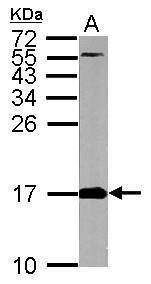
Sample (30 μg of whole cell lysate) A: Raji 15% SDS PAGE GTX111010 diluted at 1:10000 The HRP-conjugated anti-rabbit IgG antibody (GTX213110-01) was used to detect the primary antibody. |
|
|
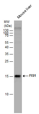
FIS1 antibody detects FIS1 protein by western blot analysis. Mouse tissue extracts (50 μg) was separated by 15% SDS-PAGE, and the membrane was blotted with FIS1 antibody (GTX111010) diluted by 1:5000. The HRP-conjugated anti-rabbit IgG antibody (GTX213110-01) was used to detect the primary antibody. |
|
|
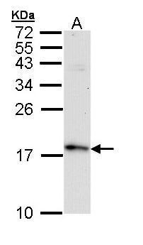
Sample (50 μg of whole cell lysate) A: mouse brain 15% SDS PAGE GTX111010 diluted at 1:5000 The HRP-conjugated anti-rabbit IgG antibody (GTX213110-01) was used to detect the primary antibody. |
|
|
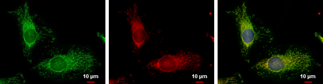
FIS1 antibody detects FIS1 protein at mitochondria by immunofluorescent analysis. Sample: HeLa cells were fixed in 2% paraformaldehyde/culture medium at 37oC for 30 min. Green: FIS1 protein stained by FIS1 antibody (GTX111010) diluted at 1:2000. Red: MitoTrackerR Red CMXRos, a mitochondria tracker. Blue: Hoechst 33342 staining. Scale bar = 10 μm. |
|
|
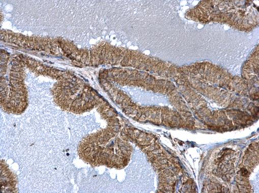
TTC11 antibody detects TTC11 protein at mitochondria on mouse prostate by immunohistochemical analysis. Sample: Paraffin-embedded mouse prostate. TTC11 antibody (GTX111010) dilution: 1:500. Antigen Retrieval: Trilogy™ (EDTA based, pH 8.0) buffer, 15min |
|
|
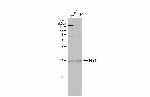
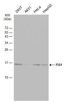
Various whole cell extracts (30 μg) were separated by 15% SDS-PAGE, and the membrane was blotted with FIS1 antibody (GTX111010) diluted at 1:10000. |
|
|
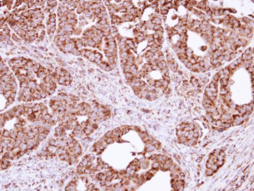
Immunohistochemical analysis of paraffin-embedded NCIN87 xenograft, using FIS1(GTX111010) antibody at 1:100 dilution. Antigen Retrieval: Trilogy™ (EDTA based, pH 8.0) buffer, 15min |
|
|
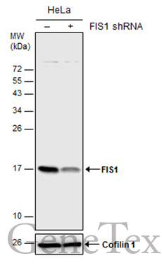
Non-transfected (–) and transfected (+) HeLa whole cell extracts (30 μg) were separated by 15% SDS-PAGE, and the membrane was blotted with FIS1 antibody (GTX111010) diluted at 1:5000. The HRP-conjugated anti-rabbit IgG antibody (GTX213110-01) was used to detect the primary antibody. |
Produktgarantie und fachkundiger Support
