
RUVBL1 antibody, Unconjugated, Rabbit, Polyclonal
Artikelnummer:
GTX111294
- Bilder (9)
| Artikelname: | RUVBL1 antibody, Unconjugated, Rabbit, Polyclonal |
| Artikelnummer: | GTX111294 |
| Hersteller Artikelnummer: | GTX111294 |
| Alternativnummer: | GTX111294-100,GTX111294-25 |
| Hersteller: | GeneTex |
| Wirt: | Rabbit |
| Kategorie: | Antikörper |
| Applikation: | ICC, IHC-P, IP, WB |
| Spezies Reaktivität: | Human, Mouse, Rat |
| Immunogen: | Recombinant protein encompassing a sequence within the center region of human RUVBL1. The exact sequence is proprietary. |
| Konjugation: | Unconjugated |
| Alternative Synonym: | RuvB like AAA ATPase 1 , ECP-54 , ECP54 , INO80H , NMP 238 , NMP238 , PONTIN , Pontin52 , RVB1 , TIH1 , TIP49 , TIP49A |
| Klonalität: | Polyclonal |
| Konzentration: | 0.32 mg/ml (Please refer to the vial label for the specific concentration.) |
| Molekulargewicht: | 50 |
| NCBI: | 8607 |
| UniProt: | Q9Y265 |
| Puffer: | 1XPBS (pH7), 1% BSA, 20% Glycerol, 0.025% ProClin 300. |
| Reinheit: | Purified by antigen-affinity chromatography. |
| Formulierung: | Liquid |
| Anwendungsbeschreibung: | WB: 1:500-1:3000. ICC/IF: 1:100-1:1000. IHC-P: 1:100-1:1000. IP: 1:100-1:500. *Optimal dilutions/concentrations should be determined by the researcher.Not tested in other applications. |
|
|
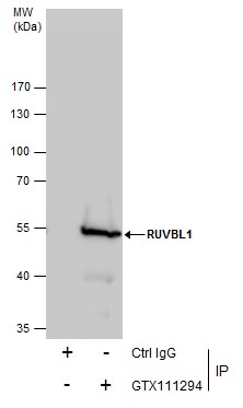
GTX111294 IP Image |
|
|
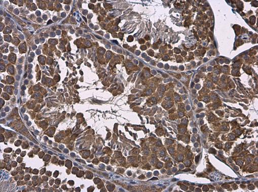
RUVBL1 antibody detects RUVBL1 protein at cytoplasm in mouse testis by immunohistochemical analysis. Sample: Paraffin-embedded mouse testis. RUVBL1 antibody (GTX111294) diluted at 1:500. Antigen Retrieval: Citrate buffer, pH 6.0, 15 min |
|
|
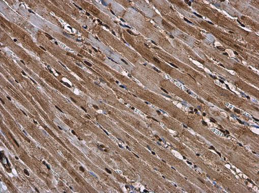
RUVBL1 antibody detects RUVBL1 protein at cytoplasm and nucleus in rat heart by immunohistochemical analysis. Sample: Paraffin-embedded rat heart. RUVBL1 antibody (GTX111294) diluted at 1:500. Antigen Retrieval: Citrate buffer, pH 6.0, 15 min |
|
|
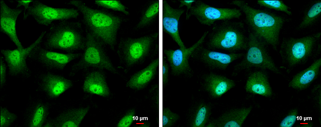
RUVBL1 antibody detects RUVBL1 protein at cytoplasm and nucleus by immunofluorescent analysis. Sample: HeLa cells were fixed in 4% paraformaldehyde at RT for 15 min. Green: RUVBL1 protein stained by RUVBL1 antibody (GTX111294) diluted at 1:500. Blue: Hoechst 33342 staining. Scale bar = 10 μm. |
|
|
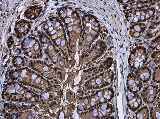
Immunohistochemical analysis of paraffin-embedded human colon carcinoma, using RUVBL1(GTX111294) antibody at 1:500 dilution. Antigen Retrieval: Trilogy™ (EDTA based, pH 8.0) buffer, 15min |
|
|
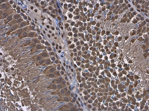
RUVBL1 antibody detects RUVBL1 protein at cytoplasm and nucleus in mouse colon by immunohistochemical analysis. Sample: Paraffin-embedded mouse colon. RUVBL1 antibody (GTX111294) diluted at 1:500. Antigen Retrieval: Citrate buffer, pH 6.0, 15 min |
|
|
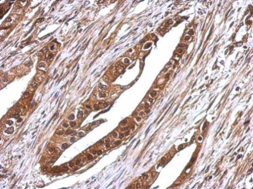
RUVBL1 antibody detects RUVBL1 protein at cytoplasm in rat testis by immunohistochemical analysis. Sample: Paraffin-embedded rat testis. RUVBL1 antibody (GTX111294) diluted at 1:500. Antigen Retrieval: Citrate buffer, pH 6.0, 15 min |
|
|
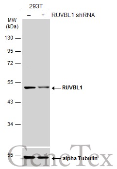
Non-transfected (–) and transfected (+) 293T whole cell extracts (30 μg) were separated by 10% SDS-PAGE, and the membrane was blotted with RUVBL1 antibody (GTX111294) diluted at 1:5000. The HRP-conjugated anti-rabbit IgG antibody (GTX213110-01) was used to detect the primary antibody. |
|
|
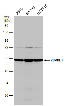
Various whole cell extracts (30 μg) were separated by 10% SDS-PAGE, and the membrane was blotted with RUVBL1 antibody (GTX111294) diluted at 1:1000. |
Produktgarantie und fachkundiger Support
