
Tyrosine Hydroxylase antibody, Unconjugated, Rabbit, Polyclonal
Artikelnummer:
GTX113016
- Bilder (9)


| Artikelname: | Tyrosine Hydroxylase antibody, Unconjugated, Rabbit, Polyclonal |
| Artikelnummer: | GTX113016 |
| Hersteller Artikelnummer: | GTX113016 |
| Alternativnummer: | GTX113016-100,GTX113016-25 |
| Hersteller: | GeneTex |
| Wirt: | Rabbit |
| Kategorie: | Antikörper |
| Applikation: | ICC, IHC-Fr, IHC-P, WB |
| Spezies Reaktivität: | Bovine, Human, Mouse, Rat, Rodent, Zebrafish |
| Immunogen: | Recombinant protein encompassing a sequence within the center region of human Tyrosine Hydroxylase. The exact sequence is proprietary. |
| Konjugation: | Unconjugated |
| Alternative Synonym: | tyrosine hydroxylase , DYT14 , DYT5b , TYH |
| Klonalität: | Polyclonal |
| Konzentration: | 0.21 mg/ml (Please refer to the vial label for the specific concentration.) |
| Molekulargewicht: | 59 |
| NCBI: | 7054 |
| UniProt: | P07101 |
| Puffer: | 1XPBS (pH7), 1% BSA, 20% Glycerol, 0.025% ProClin 300. |
| Reinheit: | Purified by antigen-affinity chromatography. |
| Formulierung: | Liquid |
| Anwendungsbeschreibung: | WB: 1:500-1:3000. ICC/IF: 1:100-1:1000. IHC-P: 1:100-1:1000. IHC-Fr: 1:100-1:1000. *Optimal dilutions/concentrations should be determined by the researcher.Not tested in other applications. |
|
|
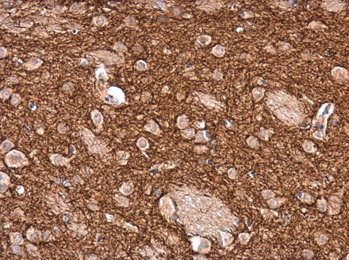
GTX113016 IHC-P Image |
|
|
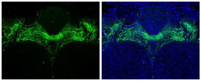
Tyrosine Hydroxylase antibody detects Tyrosine Hydroxylase protein in midbrain dopaminergic neurons by immunohistochemical analysis.Sample: Paraffin-embedded mouse brain.Green: Tyrosine Hydroxylase stained by Tyrosine Hydroxylase antibody (GTX113016) diluted at 1:1000.Blue: Fluoroshield with DAPI (GTX30920).Antigen Retrieval: Citrate buffer, pH 6.0, 15 min |
|
|
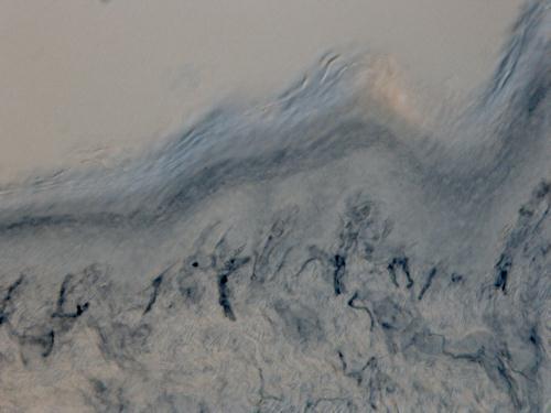
Immunohistochemical analysis of Rat hindlimb pad skin tissue (paraformaldehyde-fixed frozen sections), using Tyrosine Hydroxylase(GTX113016) antibody at 1:100 dilution. Antigen Retrieval: Citrate buffer, pH 6.0, 15 min |
|
|
Tyrosine Hydroxylase antibody detects Tyrosine Hydroxylase protein at cytoplasm by immunofluorescent analysis. Sample: U-87 MG cells were fixed in 4% paraformaldehyde at RT for 15 min. Green: Tyrosine Hydroxylase protein stained by Tyrosine Hydroxylase antibody (GTX113016) diluted at 1:400. Red: beta Tubulin 3/ TUJ1 protein stained by beta Tubulin 3/ TUJ1 antibody (GTX631836) diluted at 1:200. Blue: Hoechst 33342 staining. |
|
|
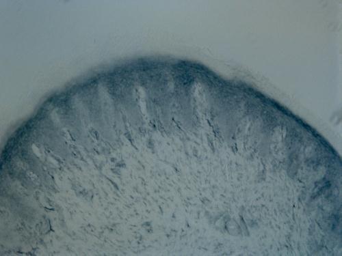
Immunohistochemical analysis of Rat hindlimb pad skin tissue (paraformaldehyde-fixed frozen sections), using Tyrosine Hydroxylase(GTX113016) antibody at 1:100 dilution. Antigen Retrieval: Citrate buffer, pH 6.0, 15 min |
|
|
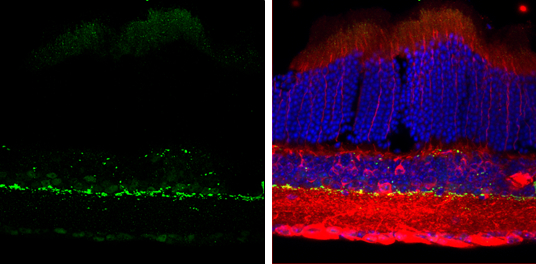
Tyrosine Hydroxylase antibody detects Tyrosine Hydroxylase protein by immunohistochemical analysis. Sample: Frozen sectioned adult mouse retina. Green: Tyrosine Hydroxylase protein stained by Tyrosine Hydroxylase antibody (GTX113016) diluted at 1:250. Red: beta Tubulin 3/ TUJ1, stained by beta Tubulin 3/ TUJ1 antibody [GT11710] (GTX631836) diluted at 1:250. Blue: Fluoroshield with DAPI (GTX30920). |
|
|
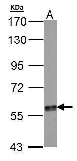
Tyrosine Hydroxylase antibody detects TH protein by western blot analysis. A. 50 μg mouse brain lysate/extract 7.5% SDS-PAGE Tyrosine Hydroxylase antibody (GTX113016) dilution: 1:500 The HRP-conjugated anti-rabbit IgG antibody (GTX213110-01) was used to detect the primary antibody. |
|
|
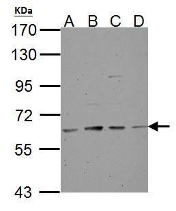
Tyrosine Hydroxylase antibody detects TH protein by western blot analysis. A. 30 μg NT2D1 whole cell lysate/extract B. 30 μg PC-3 whole cell lysate/extract C. 30 μg U87-MG whole cell lysate/extract D. 30 μg SK-N-SH whole cell lysate/extract 7.5% SDS-PAGE Tyrosine Hydroxylase antibody (GTX113016) dilution: 1:500 The HRP-conjugated anti-rabbit IgG antibody (GTX213110-01) was used to detect the primary antibody. |
|
|
Rat tissue extract (50 μg) was separated by 7.5% SDS-PAGE, and the membrane was blotted with Tyrosine Hydroxylase antibody (GTX113016) diluted at 1:1000. |
Produktgarantie und fachkundiger Support
