
ROCK1 antibody [N1N2], N-term, Unconjugated, Rabbit, Polyclonal
Artikelnummer:
GTX113266
- Bilder (9)

| Artikelname: | ROCK1 antibody [N1N2], N-term, Unconjugated, Rabbit, Polyclonal |
| Artikelnummer: | GTX113266 |
| Hersteller Artikelnummer: | GTX113266 |
| Alternativnummer: | GTX113266-100,GTX113266-25 |
| Hersteller: | GeneTex |
| Wirt: | Rabbit |
| Kategorie: | Antikörper |
| Applikation: | ICC, IHC-P, IP, WB |
| Spezies Reaktivität: | Human, Mouse, Rat |
| Immunogen: | Recombinant protein encompassing a sequence within the N-terminus region of human ROCK1. The exact sequence is proprietary. |
| Konjugation: | Unconjugated |
| Alternative Synonym: | Rho associated coiled-coil containing protein kinase 1 , P160ROCK , ROCK-I |
| Anwendungsbeschreibung: | WB: 1:500-1:3000. ICC/IF: 1:100-1:1000. IHC-P: 1:100-1:1000. IP: 1:100-1:500. *Optimal dilutions/concentrations should be determined by the researcher.Not tested in other applications. |
|
|
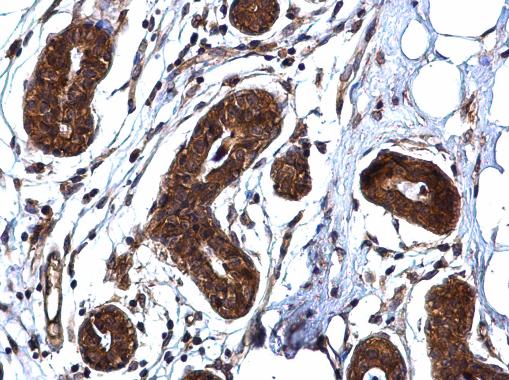
GTX113266 IHC-P Image |
|
|
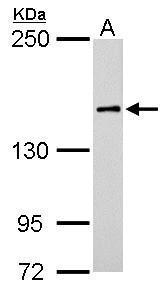
ROCK1 antibody [N1N2], N-term detects ROCK1 protein by western blot analysis. A. 50 μg rat brain lysate/extract 5% SDS-PAGE ROCK1 antibody [N1N2], N-term (GTX113266) dilution: 1:500 The HRP-conjugated anti-rabbit IgG antibody (GTX213110-01) was used to detect the primary antibody. |
|
|
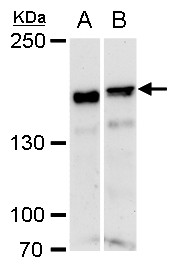
ROCK1 antibody [N1N2], N-term detects ROCK1 protein by western blot analysis. A. 30 μg NIH-3T3 whole cell extract B. 30 μg C2Cl2 whole cell extract 5% SDS-PAGE ROCK1 antibody [N1N2], N-term (GTX113266) dilution: 1:1000 The HRP-conjugated anti-rabbit IgG antibody (GTX213110-01) was used to detect the primary antibody. |
|
|
ROCK1 antibody immunoprecipitates ROCK1 protein in IP experiments. IP Sample: A431 whole cell lysate/extract A : Control with 3 μg of pre-immune rabbit IgG B : Immunoprecipitation of ROCK1 by 3 μg of ROCK1 antibody (GTX113266) 5% SDS-PAGE The immunoprecipitated ROCK1 protein was detected by ROCK1 antibody (GTX113266) diluted at 1 : 500. EasyBlot anti-rabbit IgG (HRP) (GTX221666-01) was used as a secondary reagent. |
|
|
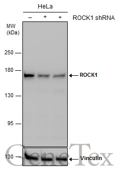
Non-transfected (–) and transfected (+) HeLa whole cell extracts (30 μg) were separated by 5% SDS-PAGE, and the membrane was blotted with ROCK1 antibody [N1N2], N-term (GTX113266) diluted at 1:1000. The HRP-conjugated anti-rabbit IgG antibody (GTX213110-01) was used to detect the primary antibody. |
|
|
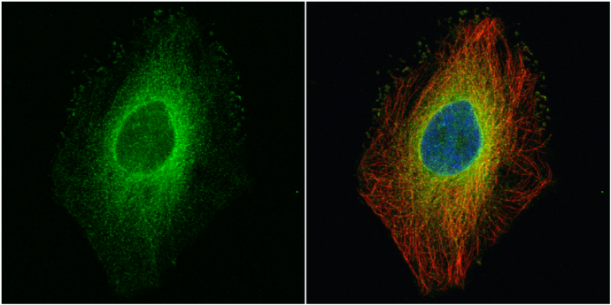
ROCK1 antibody [N1N2], N-term detects ROCK1 protein at cytoplasm by immunofluorescent analysis. Sample: HeLa cells were fixed in 4% paraformaldehyde at RT for 15 min. Green: ROCK1 protein stained by ROCK1 antibody [N1N2], N-term (GTX113266) diluted at 1:500. Red: alpha Tubulin, a cytoskeleton marker, stained by alpha Tubulin antibody [GT114] (GTX628802) diluted at 1:500. Blue: Hoechst 33342 staining. |
|
|
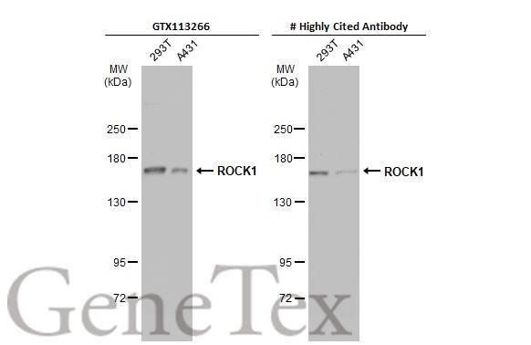
Various whole cell extracts (30 μg) were separated by 5% SDS-PAGE, and the membranes were blotted with ROCK1 antibody [N1N2], N-term (GTX113266) diluted at 1:1000 and competitor's antibody diluted at 1:1000. The HRP-conjugated anti-rabbit IgG antibody (GTX213110-01) was used to detect the primary antibody, and the signal was developed with Trident ECL plus-Enhanced. *The competitor is not affiliated with GeneTex and does not endorse this product. |
|
|
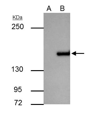
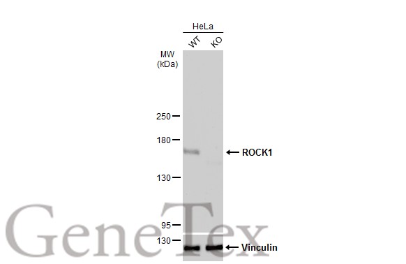
Wild-type (WT) and ROCK1 knockout (KO) HeLa cell extracts (30 μg) were separated by 5% SDS-PAGE, and the membrane was blotted with ROCK1 antibody [N1N2], N-term (GTX113266) diluted at 1:1000. The HRP-conjugated anti-rabbit IgG antibody (GTX213110-01) was used to detect the primary antibody. |
|
|
Various whole cell extracts (30 μg) were separated by 5% SDS-PAGE, and the membrane was blotted with ROCK1 antibody [N1N2], N-term (GTX113266) diluted at 1:1000. The HRP-conjugated anti-rabbit IgG antibody (GTX213110-01) was used to detect the primary antibody. |
Produktgarantie und fachkundiger Support
