
p53 antibody, Unconjugated, Rabbit, Polyclonal
Artikelnummer:
GTX50438
- Bilder (9)
| Artikelname: | p53 antibody, Unconjugated, Rabbit, Polyclonal |
| Artikelnummer: | GTX50438 |
| Hersteller Artikelnummer: | GTX50438 |
| Alternativnummer: | GTX50438-100 |
| Hersteller: | GeneTex |
| Wirt: | Rabbit |
| Kategorie: | Antikörper |
| Applikation: | ICC, IHC-P, IP, WB |
| Spezies Reaktivität: | Human, Mouse, Rat |
| Immunogen: | Peptide sequence around aa.13~17 (P-L-S-Q-E) derived from human p53. |
| Konjugation: | Unconjugated |
| Alternative Synonym: | tumor protein p53 , BCC7 , BMFS5 , LFS1 , P53 , TRP53 |
| This gene encodes a tumor suppressor protein containing transcriptional activation, DNA binding, and oligomerization domains. The encoded protein responds to diverse cellular stresses to regulate expression of target genes, thereby inducing cell cycle ar |
| Klonalität: | Polyclonal |
| Konzentration: | 1 mg/ml (Please refer to the vial label for the specific concentration.) |
| Klon-Bezeichnung: | Polyclonal |
| Molekulargewicht: | 44 |
| Isotyp: | IgG |
| NCBI: | 7157 |
| Pubmed: | 32890980, |
| UniProt: | P04637 |
| Puffer: | PBS (without Mg and Ca) pH7.4, 150mM NaCl, 50% Glycerol, 0.02% Sodium azide. |
| Quelle: | Human |
| Reinheit: | Purified by antigen-affinity chromatography. |
| Formulierung: | Liquid |
| Anwendungsbeschreibung: | WB: 1:500-1:1000. ICC/IF: 1:100-1:200. IHC-P: 1:50-1:100. *Optimal dilutions/concentrations should be determined by the researcher.Not tested in other applications. |
|
|
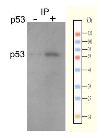
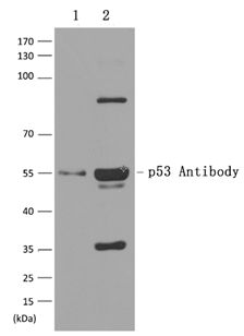
IP analysis of extracts from 293T cells using GTX50438 p53 antibody. |
|
|
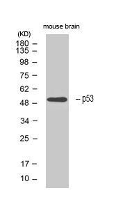
WB analysis of extracts from mouse brain tissue using GTX50438 p53 antibody. |
|
|
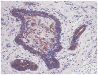
IHC-P analysis of rat breast tissue sections using GTX50438 p53 antibody. |
|
|
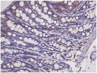
IHC-P analysis of rat colon tissue sections using GTX50438 p53 antibody. |
|
|
IHC-P analysis of rat breast tissue using GTX50438 p53 antibody. Dilution : 1:100 |
|
|
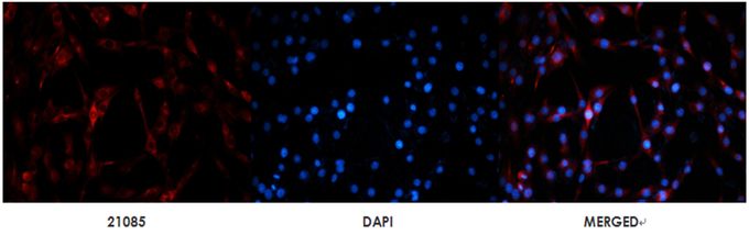
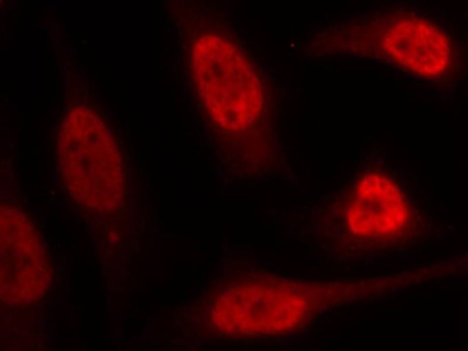
ICC/IF analysis of methanol-fixed HeLa cells using GTX50438 p53 antibody. |
|
|
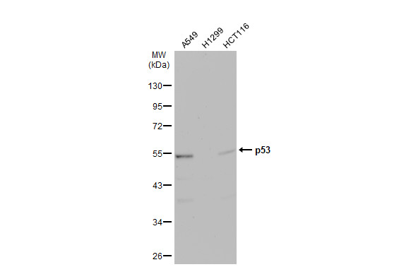
GTX50438 WB Image |
|
|
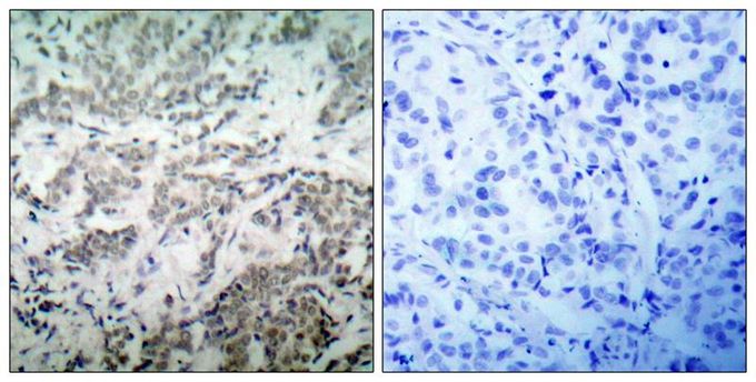
IHC-P analysis of human breast carcinoma tissue using GTX50438 p53 antibody. Left : Primary antibody Right : Primary antibody pre-incubated with the antigen specific peptide |
|
|
Various whole cell extracts (30 μg) were separated by 10% SDS-PAGE, and the membrane was blotted with p53 antibody (GTX50438) diluted at 1:500. The HRP-conjugated anti-rabbit IgG antibody (GTX213110-01) was used to detect the primary antibody, and the signal was developed with Trident ECL plus-Enhanced. |
Produktgarantie und fachkundiger Support
