
NDP52 antibody [GT422], IgG1, Unconjugated, Mouse, Monoclonal
Artikelnummer:
GTX630396
- Bilder (9)
| Artikelname: | NDP52 antibody [GT422], IgG1, Unconjugated, Mouse, Monoclonal |
| Artikelnummer: | GTX630396 |
| Hersteller Artikelnummer: | GTX630396 |
| Alternativnummer: | GTX630396-100,GTX630396-25 |
| Hersteller: | GeneTex |
| Wirt: | Mouse |
| Kategorie: | Antikörper |
| Applikation: | ICC, IP, WB |
| Spezies Reaktivität: | Human |
| Immunogen: | Recombinant protein encompassing a sequence within the center region of human NDP52. The exact sequence is proprietary. |
| Konjugation: | Unconjugated |
| Alternative Synonym: | calcium binding and coiled-coil domain 2 , NDP52 |
| Anwendungsbeschreibung: | WB: 1:500-1:3000. ICC/IF: 1:100-1:1000. IP: 1:100-1:500. *Optimal dilutions/concentrations should be determined by the researcher.Not tested in other applications. |
|
|
GTX630396 WB Image |
|
|
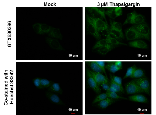
NDP52 antibody [GT422] detects NDP52 protein at autophagosome by immunofluorescent analysis. Samples: Hep G2 cells mock (left) and treated with 3 μM Thapsigargin for 16 hrs (right) were fixed in ice-cold MeOH for 10 min and permeabilized with 100% MeOH for 30 sec. Green: NDP52 protein stained by NDP52 antibody [GT422] (GTX630396) diluted at 1:1000. Blue: Hoechst 33342 staining. Scale bar = 10 μm. |
|
|
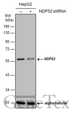
Non-transfected (–) and transfected (+) HepG2 whole cell extracts (30 μg) were separated by 10% SDS-PAGE, and the membrane was blotted with NDP52 antibody [GT422] (GTX630396) diluted at 1:1000. |
|
|
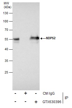
Immunoprecipitation of NDP52 protein from Jurkat whole cell extracts using 5 μg of NDP52 antibody [GT422] (GTX630396). Western blot analysis was performed using NDP52 antibody [GT422] (GTX630396). EasyBlot anti-Mouse IgG (GTX221667-01) was used as a secondary reagent. |
|
|
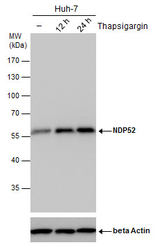
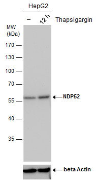
NDP52 antibody detects NDP52 protein by western blot analysis. Un-treated (-) and treated (+, Thapsigargin treatment for 12hrs) HepG2 whole cell extracts (30 μg) were separated by 10% SDS-PAGE, and the membrane was blotted with NDP52 antibody (GTX630396) diluted by 1:500. The ACTB was used as internal control (GTX110564, 1:50000) shown at the bottom panel. |
|
|
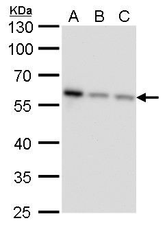
NDP52 antibody [GT422] detects NDP52 protein by western blot analysis. A. 30 μg Jurkat whole cell lysate/extract B. 30 μg Raji whole cell lysate/extract C. 30 μg NCI-H929 whole cell lysate/extract 10 % SDS-PAGE NDP52 antibody [GT422] (GTX630396) dilution: 1:1000 |
|
|
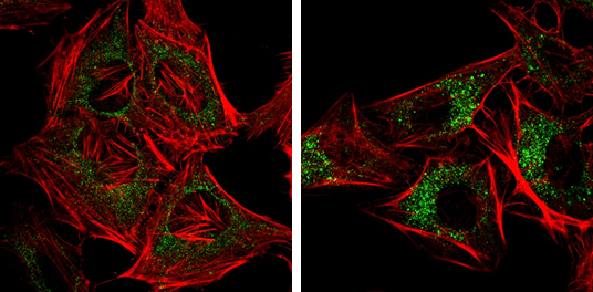
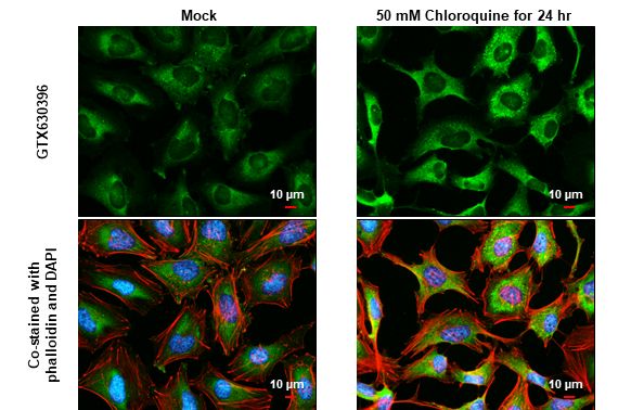
NDP52 antibody [GT422] detects NDP52 protein at autophagosome by immunofluorescent analysis. Samples: HeLa cells mock (left) and treated with 50μM Chloroquine for 24 hr (right) were fixed in 4% paraformaldehyde at RT for 15 min. Green: NDP52 protein stained by NDP52 antibody [GT422] (GTX630396) diluted at 1:1000. Red: Phalloidin, a F-actin marker. |
|
|
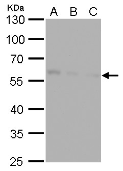
NDP52 antibody [GT422] detects NDP52 protein by western blot analysis. A. 30 μg Huh7 whole cell lysate/extract B. 30 μg Hep3B whole cell lysate/extract C. 30 μg HepG2 whole cell lysate/extract 10 % SDS-PAGE NDP52 antibody [GT422] (GTX630396) dilution: 1:1000 |
|
|
NDP52 antibody [GT422] detects NDP52 protein at autophagosome? by immunofluorescent analysis.Sample: Mock and treated HeLa cells were fixed in 4% paraformaldehyde at RT for 15 min.Green: NDP52 stained by NDP52 antibody [GT422] (GTX630396) diluted at 1:1000.Red: phalloidin, a cytoskeleton marker, diluted at 1:200.Blue: Fluoroshield with DAPI (GTX30920).Scale bar= 10 μm. |
Produktgarantie und fachkundiger Support
