
Profilin 1 antibody [2D2], IgG1, Unconjugated, Mouse, Monoclonal
Artikelnummer:
GTX83902
- Bilder (9)
| Artikelname: | Profilin 1 antibody [2D2], IgG1, Unconjugated, Mouse, Monoclonal |
| Artikelnummer: | GTX83902 |
| Hersteller Artikelnummer: | GTX83902 |
| Alternativnummer: | GTX83902-100 |
| Hersteller: | GeneTex |
| Wirt: | Mouse |
| Kategorie: | Antikörper |
| Applikation: | FACS, ICC, IHC-P, WB |
| Spezies Reaktivität: | Canine, Human, Monkey, Rat |
| Immunogen: | Full length human recombinant protein of human PFN1 (NP_005013) produced in HEK293T cell. |
| Konjugation: | Unconjugated |
| Alternative Synonym: | profilin 1 , ALS18 |
| Binds to actin and affects the structure of the cytoskeleton. At high concentrations, profilin prevents the polymerization of actin, whereas it enhances it at low concentrations. By binding to PIP2, it inhibits the formation of IP3 and DG. |
| Klonalität: | Monoclonal |
| Konzentration: | 0.58 mg/ml (Please refer to the vial label for the specific concentration.) |
| Klon-Bezeichnung: | [2D2] |
| Molekulargewicht: | 15 |
| Isotyp: | IgG1 |
| NCBI: | 5216 |
| UniProt: | P07737 |
| Puffer: | PBS pH7.3, 1% BSA, 50% Glycerol, 0.02% Sodium azide. |
| Quelle: | Human |
| Reinheit: | Purified by affinity chromatography |
| Formulierung: | Liquid |
| Anwendungsbeschreibung: | WB: 1:2000. ICC/IF: 1:100. IHC-P: 1:50. FACS: 1:100. *Optimal dilutions/concentrations should be determined by the researcher.Not tested in other applications. |
|
|
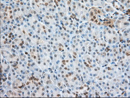
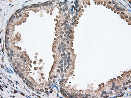
IHC-P analysis of human pancreas tissue using GTX83902 Profilin 1 antibody [2D2]. Antigen retrieval : Heat-induced epitope retrieval by 10mM citrate buffer, pH6.0, 100ºC for 10min. Dilution : 1:50 |
|
|
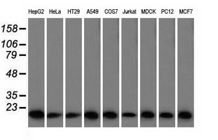
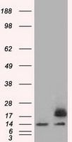
WB analysis of various cell lines using GTX83902 Profilin 1 antibody [2D2]. Loading : 35 ug per lane |
|
|
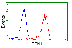
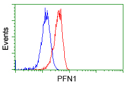
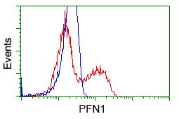
FACS analysis of Jurkat cells using GTX83902 Profilin 1 antibody [2D2]. Red : Primary antibody Blue : Negative control antibody |
|
|
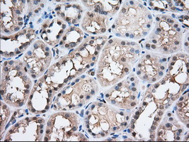
IHC-P analysis of human kidney tissue using GTX83902 Profilin 1 antibody [2D2]. Antigen retrieval : Heat-induced epitope retrieval by 10mM citrate buffer, pH6.0, 100ºC for 10min. Dilution : 1:50 |
|
|
IHC-P analysis of human prostate tissue using GTX83902 Profilin 1 antibody [2D2]. Antigen retrieval : Heat-induced epitope retrieval by 10mM citrate buffer, pH6.0, 100ºC for 10min. Dilution : 1:50 |
|
|
FACS analysis of HeLa cells using GTX83902 Profilin 1 antibody [2D2]. Red : Primary antibody Blue : Negative control antibody |
|
|
GTX83902 FACS Image |
|
|
WB analysis of HEK293T cells transfected with Profilin 1 plasmid (Right) or empty vector (Left) for 48 hrs using GTX83902 Profilin 1 antibody [2D2]. Loading : 5 ug per lane |
|
|
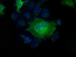
ICC/IF analysis of COS7 cells transiently transfected with Profilin 1 plasmid using GTX83902 Profilin 1 antibody [2D2]. |
Produktgarantie und fachkundiger Support
