
IMPDH2 Antibody, Clone: [JE40-87], Unconjugated, Rabbit, Recombinant
Catalog Number:
BYT-ORB622725
- Images (9)
| Article Name: | IMPDH2 Antibody, Clone: [JE40-87], Unconjugated, Rabbit, Recombinant |
| Biozol Catalog Number: | BYT-ORB622725 |
| Supplier Catalog Number: | orb622725 |
| Alternative Catalog Number: | BYT-ORB622725-100,BYT-ORB622725-50 |
| Manufacturer: | Biorbyt |
| Host: | Rabbit |
| Category: | Antikörper |
| Application: | FC, ICC, IF, IHC, IP, WB |
| Species Reactivity: | Human, Mouse, Rat |
| Immunogen: | Recombinant protein within C-terminal human IMPDH2. |
| Conjugation: | Unconjugated |
| Alternative Names: | IMDH2_HUMAN antibody/ IMP (inosine monophosphate) dehydrogenase 2 antibody/ IMP dehydrogenase 2 antibody/ IMP oxireductase 2 antibody/ IMPD 2 antibody/ IMPD2 antibody/ IMPDH 2 antibody/ IMPDH II antibody/ IMPDH-II antibody/ Impdh2 antibody/ Inosine 5 monophosphate dehydrogenase 2 antibody/ Inosine 5 phosphate dehydrogenase 2 antibody/ Inosine monophosphate dehydrogenase type II antibody/ Inosine-5-monophosphate dehydrogenase 2 antibody |
| IMPDH2 Antibody |
| Clonality: | Recombinant |
| Clone Designation: | [JE40-87] |
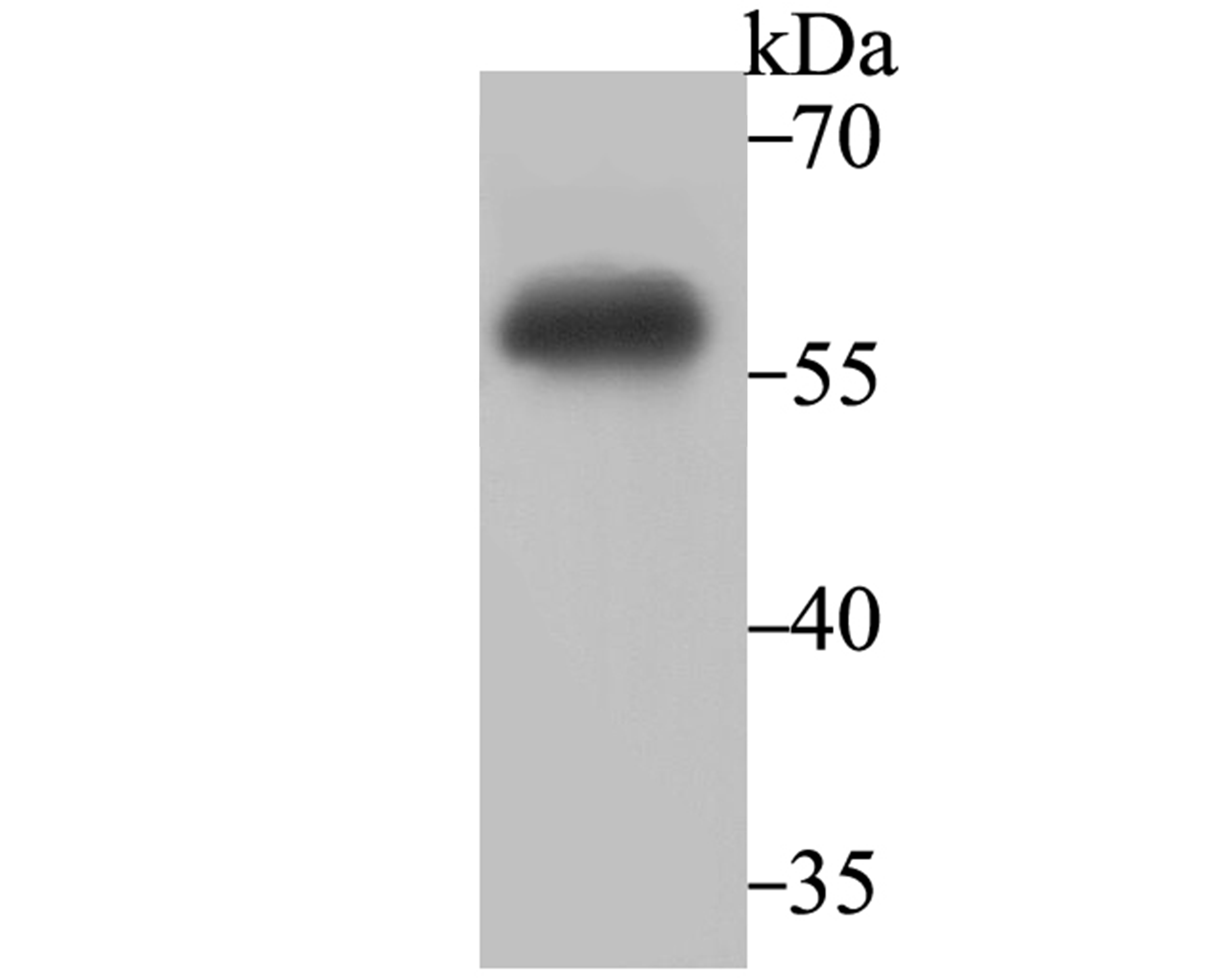
| Molecular Weight: | Calculated MW 56 kDa. |
| UniProt: | P12268 |
| Buffer: | 1*TBS (pH7.4), 1% rAlbumin, 40% Glycerol 0.05% Sodium Azide |
| Purity: | ProA affinity purified. |
| Form: | Liquid |
| Application Dilute: | WB: 1:500-1:2,000 FC: 1:10-1:50 IP: 1:10-1:50 ICC/IF: 1:50-1:200 IHC: 1:50-1:200 |
|
|
Western blot analysis of IMPDH2 on Daudi cell lysate using anti-IMPDH2 antibody at 1/2000 dilution |
|
|
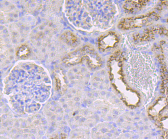
Immunohistochemical analysis of paraffin-embedded rat kidney tissue using anti-IMPDH2 antibody. Counter stained with hematoxylin |
|
|
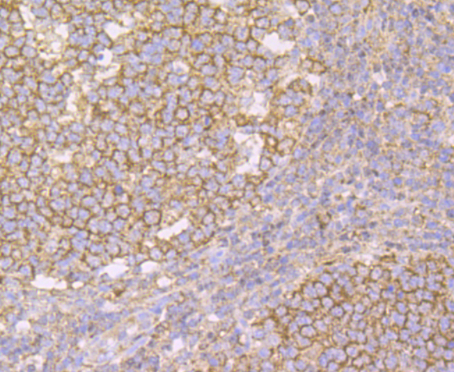
Immunohistochemical analysis of paraffin-embedded human tonsil tissue using anti-IMPDH2 antibody. Counter stained with hematoxylin |
|
|
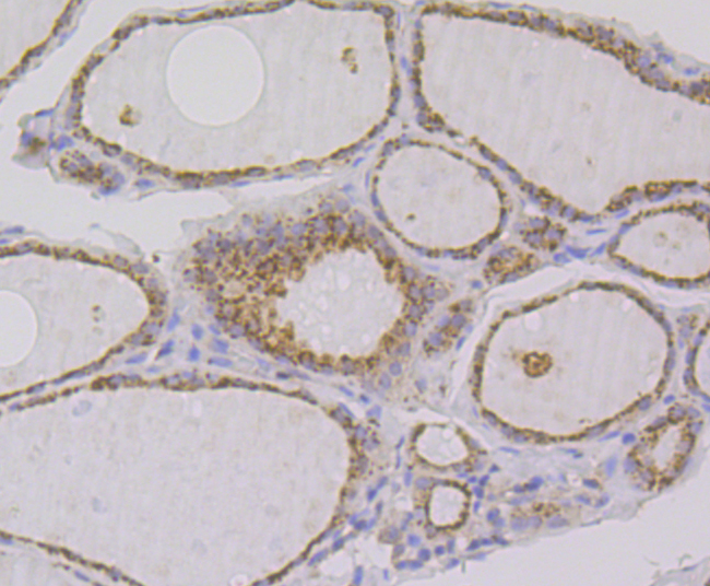
Immunohistochemical analysis of paraffin-embedded human thyroid gland tissue using anti-IMPDH2 antibody. Counter stained with hematoxylin |
|
|
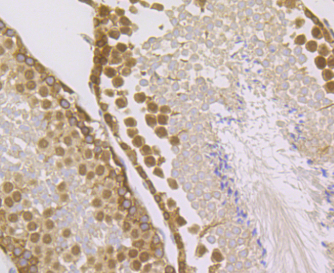
Immunohistochemical analysis of paraffin-embedded mouse testis tissue using anti-IMPDH2 antibody. Counter stained with hematoxylin |
|
|
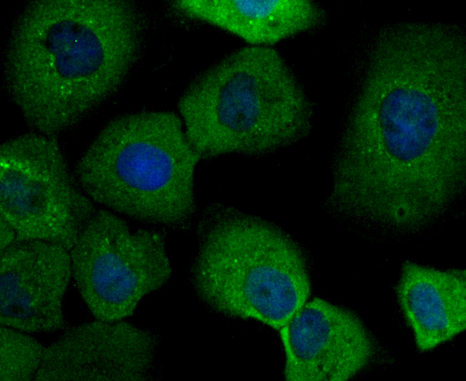
ICC staining IMPDH2 in A431 cells (green). The nuclear counter stain is DAPI (blue). Cells were fixed in paraformaldehyde, permeabilised with 0.25% Triton X100/PBS |
|
|
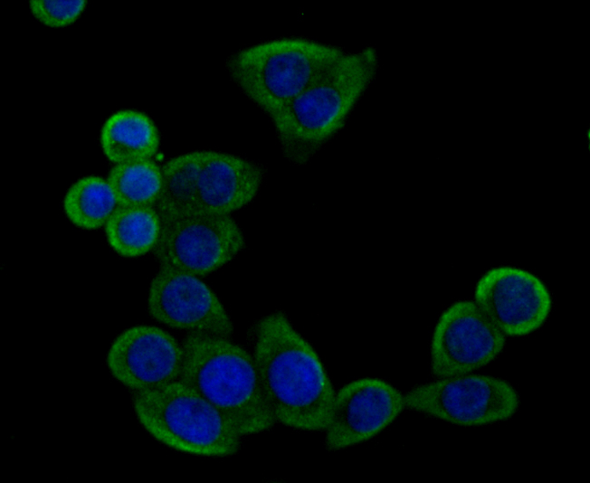
ICC staining IMPDH2 in LOVO cells (green). The nuclear counter stain is DAPI (blue). Cells were fixed in paraformaldehyde, permeabilised with 0.25% Triton X100/PBS |
|
|
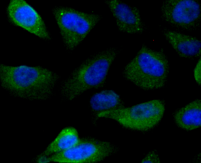
ICC staining IMPDH2 in PC-3M cells (green). The nuclear counter stain is DAPI (blue). Cells were fixed in paraformaldehyde, permeabilised with 0.25% Triton X100/PBS |
|
|
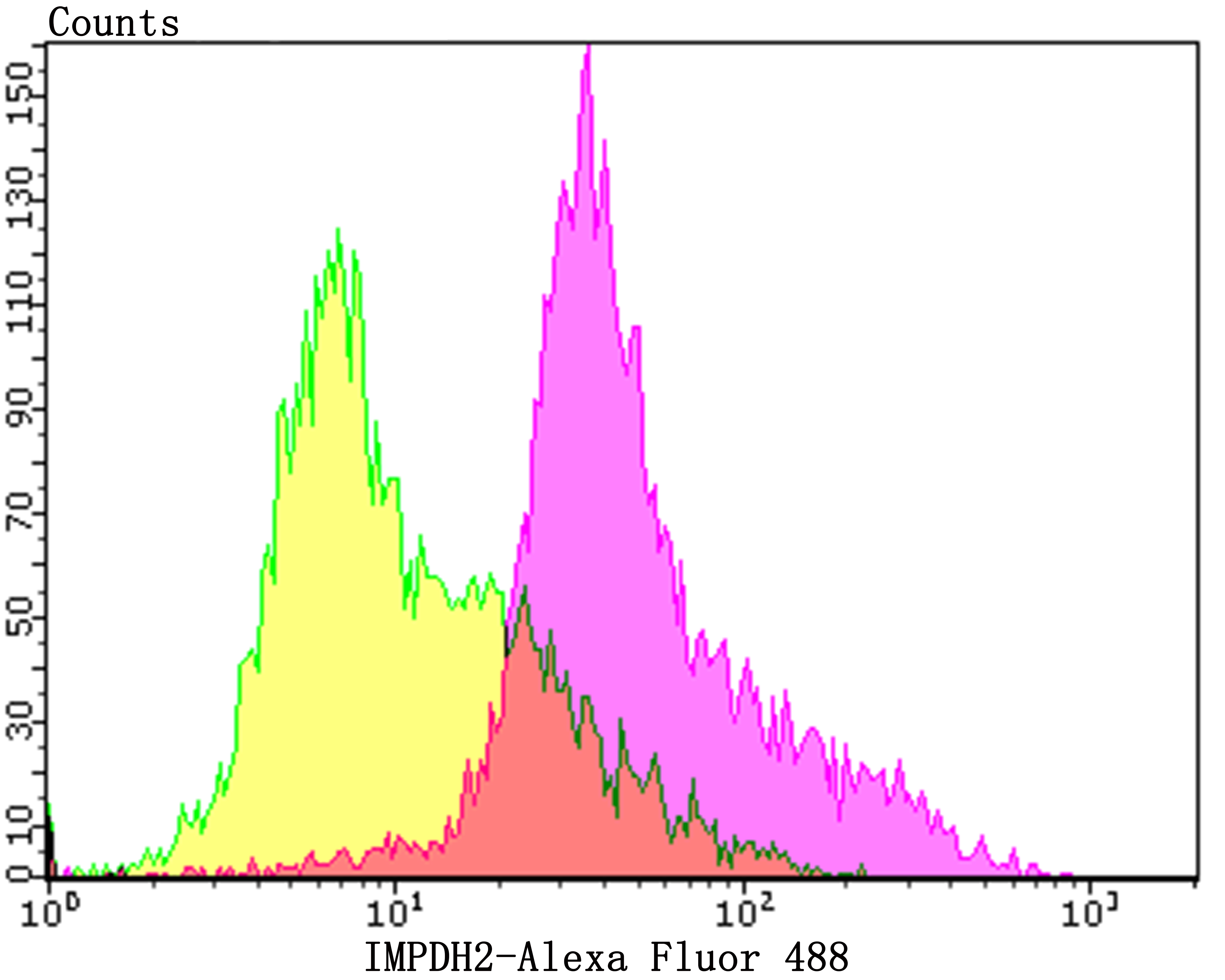
Flow cytometric analysis of Daudi cells with IMPDH2 antibody at 1/100 dilution (purple) compared with an unlabelled control (cells without incubation with primary antibody, yellow). Alexa Fluor 488-conjugated goat anti-rabbit IgG was used as the secondary antibody |
Product Guarantee and Expert Support
