
p53 antibody, Unconjugated, Rabbit, Polyclonal
Catalog Number:
GTX102965
- Images (9)
| Article Name: | p53 antibody, Unconjugated, Rabbit, Polyclonal |
| Biozol Catalog Number: | GTX102965 |
| Supplier Catalog Number: | GTX102965 |
| Alternative Catalog Number: | GTX102965-100,GTX102965-25 |
| Manufacturer: | GeneTex |
| Host: | Rabbit |
| Category: | Antikörper |
| Application: | ChIP, ICC, IHC-P, IP, WB |
| Species Reactivity: | Human, Mouse, Zebrafish |
| Immunogen: | Recombinant protein encompassing a sequence within the center region of human p53. The exact sequence is proprietary. |
| Conjugation: | Unconjugated |
| Alternative Names: | tumor protein p53 , BCC7 , BMFS5 , LFS1 , P53 , TRP53 |
| Application Notes: | WB: 1:500-1:3000. ICC/IF: 1:100-1:1000. IHC-P: 1:100-1:1000. IP: 1:100-1:500. *Optimal dilutions/concentrations should be determined by the researcher.Not tested in other applications. |
|
|
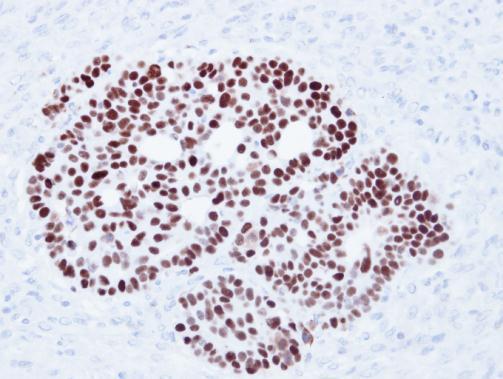
GTX102965 IHC-P Image |
|
|
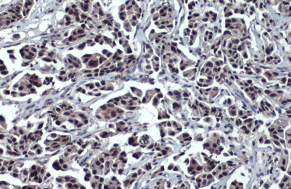
Immunohistochemical analysis of paraffin-embedded Cal27 xenograft, using p53(GTX102965) antibody at 1:500 dilution. Antigen Retrieval: Trilogy™ (EDTA based, pH 8.0) buffer, 15min |
|
|
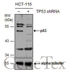
Non-transfected (–) and transfected (+) HCT116 whole cell extracts (30 μg) were separated by 10% SDS-PAGE, and the membrane was blotted with p53 antibody (GTX102965) diluted at 1:1000. The HRP-conjugated anti-rabbit IgG antibody (GTX213110-01) was used to detect the primary antibody. |
|
|
Immunoprecipitation of p53 protein. HCT116 lysates with 30uM cisplatin treatment for 24 hours were subjected to immunoprecipitation using (B) normal rabbit IgG or (C) 2.5 ug of anti-p53 antibody (GTX102965).?(A) Input, 20ug of HCT116 lysates. The precipitated protein was detected by GTX102965 diluted at 1:10000. EasyBlot anti-Rabbit IgG Kit (GTX225856-01) was used in Western blot. |
|
|
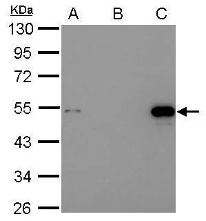
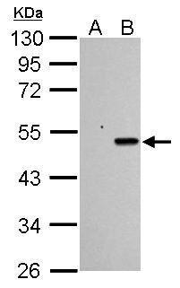
Sample (30 μg of whole cell lysate) A: HCT116 cells with mock treatment for 24 hr B: HCT116 cells with 30 μM cisplatin treatment for 24 hr 10% SDS PAGE GTX102965 diluted at 1:1000 The HRP-conjugated anti-rabbit IgG antibody (GTX213110-01) was used to detect the primary antibody. |
|
|
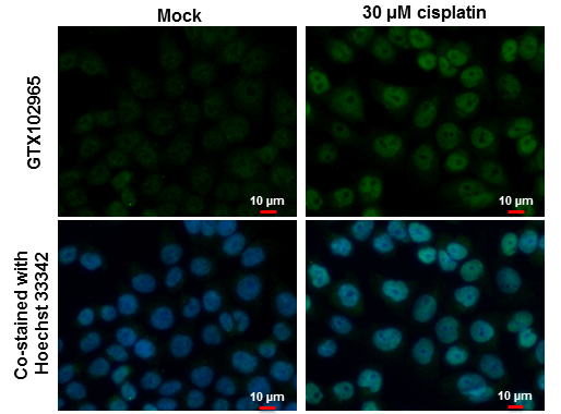
p53 antibody detects p53 protein at nucleus by immunofluorescent analysis. Samples: HCT 116 cells mock (left) and treated with 30 μM Cisplatin for 24 hrs (right) were fixed in 4% paraformaldehyde at RT for 15 min. Green: p53 protein stained by p53 antibody (GTX102965) diluted at 1:500. Blue: Hoechst 33342 staining. Scale bar = 10 μm. |
|
|
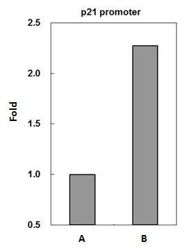
p53 antibody immunoprecipitates p53 protein-DNA complex in ChIP experiments. ChIP Sample: HCT116 whole cell lysate/extract treated with CPT 500nM for 6hr A. 5 μg preimmune rabbit IgG B. 5 μg of p53 antibody (GTX102965) The precipitated DNA was detected by PCR with primer set targeting to p21 promoter. |
|
|
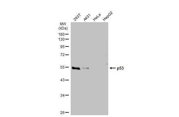
Various whole cell extracts (30 μg) were separated by 10% SDS-PAGE, and the membrane was blotted with p53 antibody (GTX102965) diluted at 1:1000. The HRP-conjugated anti-rabbit IgG antibody (GTX213110-01) was used to detect the primary antibody. |
|
|
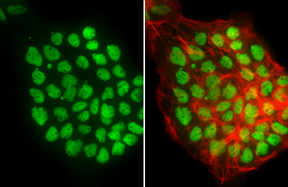
p53 antibody detects p53 protein at nucleus by immunofluorescent analysis.Sample: A431 cells were fixed in 4% paraformaldehyde at RT for 15 min.Green: p53 stained by p53 antibody (GTX102965) diluted at 1:500.Red: phalloidin, a cytoskeleton marker, diluted at 1:200. |
Product Guarantee and Expert Support
