
SOX2 antibody [GT1352], IgG2a, Unconjugated, Mouse, Monoclonal
Catalog Number:
GTX627405
- Images (9)
| Article Name: | SOX2 antibody [GT1352], IgG2a, Unconjugated, Mouse, Monoclonal |
| Biozol Catalog Number: | GTX627405 |
| Supplier Catalog Number: | GTX627405 |
| Alternative Catalog Number: | GTX627405-100,GTX627405-25 |
| Manufacturer: | GeneTex |
| Host: | Mouse |
| Category: | Antikörper |
| Application: | FACS, ICC, IHC-Fr, IHC-P, WB |
| Species Reactivity: | Human, Mouse |
| Immunogen: | Recombinant protein encompassing a sequence within the center region of human SOX2. The exact sequence is proprietary. |
| Conjugation: | Unconjugated |
| Alternative Names: | SRY-box 2 , ANOP3 , MCOPS3 |
| Application Notes: | WB: 1:500-1:3000. ICC/IF: 1:100-1:1000. IHC-P: 1:100-1:1000. IHC-Fr: 1:100-1:1000. FACS: 1:50-1:200. *Optimal dilutions/concentrations should be determined by the researcher.Not tested in other applications. |
|
|
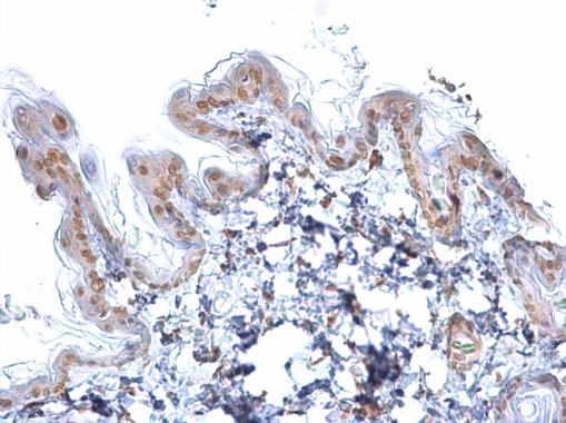
GTX627405 IHC-P Image |
|
|
SOX2 antibody [GT1352] detects SOX2 protein at nucleus by immunohistochemical analysis.Sample: Paraffin-embedded mouse E10.5 mouse embryo.Green: Brachyury stained by Brachyury antibody (GTX133714) diluted at 1:250.Red: SOX2, a nucleus marker, stained by SOX2 antibody [GT1352] (GTX627405) diluted at 1:250.Blue: Fluoroshield with DAPI (GTX30920). |
|
|
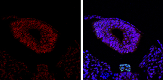
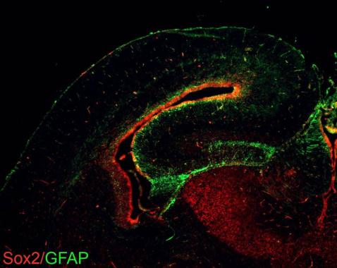
Sox2 antibodies detects Sox2 proteins on embryonic mouse brain by immunohistochemical analysis. Sample: Frozen section of embryonic mouse brain (mE18.5). Green: GFAP antibody (GTX108711) diluted at 1:1000. Red: Sox2 antibody [GT1352] (GTX627405) diluted at 1:250. |
|
|
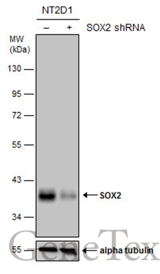
Non-transfected (–) and transfected (+) NT2D1 whole cell extracts (30 μg) were separated by 10% SDS-PAGE, and the membrane was blotted with SOX2 antibody [GT1352] (GTX627405) diluted at 1:500. The HRP-conjugated anti-mouse IgG antibody (GTX213111-01) was used to detect the primary antibody. |
|
|
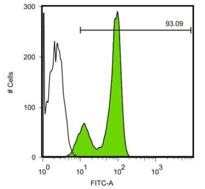
SOX2 antibody [GT1352] detects SOX2 protein by flow cytomertry analysis. Sample: Human embryonic stem cells Black: Isotype control dilution: 1:50 Green: SOX2 antibody [GT1352] dilution: 1:50 |
|
|
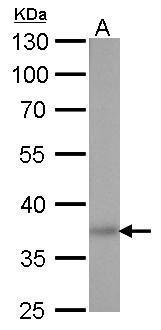
SOX2 antibody [GT1352] detects SOX2 protein by western blot analysis. A. 30 μg human ESC whole cell lysate/extract 10% SDS-PAGE SOX2 antibody [GT1352] (GTX627405) dilution: 1:1000 The HRP-conjugated anti-mouse IgG antibody (GTX213111-01) was used to detect the primary antibody. |
|
|
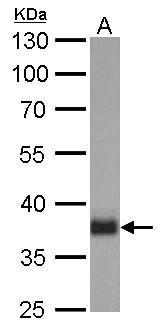
SOX2 antibody [GT1352] detects SOX2 protein by western blot analysis. A. 30 μg mouse ESC whole cell lysate/extract 10% SDS-PAGE SOX2 antibody [GT1352] (GTX627405) dilution: 1:1000 The HRP-conjugated anti-mouse IgG antibody (GTX213111-01) was used to detect the primary antibody. |
|
|
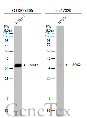
Whole cell extract (30 μg) was separated by 12% SDS-PAGE, and the membranes were blotted with SOX2 antibody [GT1352] (GTX627405) diluted at 1:1000 and competitor's antibody (sc-17320) diluted at 1:500. The HRP-conjugated anti-mouse IgG antibody (GTX213111-01) was used to detect the primary antibody, and the signal was developed with Trident ECL plus-Enhanced. *The competitor is not affiliated with GeneTex and does not endorse this product. |
|
|
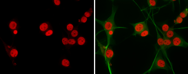
SOX2 antibody detects SOX2 protein at nucleus by immunofluorescent analysis. Sample: U-87 MG cells were fixed in 4% paraformaldehyde at RT for 15 min. Red: SOX2 protein stained by SOX2 antibody (GTX627405) diluted at 1:200. Green: beta Tubulin 3/ TUJ1 protein stained by beta Tubulin 3/ TUJ1 antibody (GTX631836) diluted at 1:200. |
Product Guarantee and Expert Support
