
EGFR antibody [GT133], IgG2b, Unconjugated, Mouse, Monoclonal
Catalog Number:
GTX628887
- Images (9)
| Article Name: | EGFR antibody [GT133], IgG2b, Unconjugated, Mouse, Monoclonal |
| Biozol Catalog Number: | GTX628887 |
| Supplier Catalog Number: | GTX628887 |
| Alternative Catalog Number: | GTX628887-100,GTX628887-25 |
| Manufacturer: | GeneTex |
| Host: | Mouse |
| Category: | Antikörper |
| Application: | ICC, IHC-P, IP, WB |
| Species Reactivity: | Human |
| Immunogen: | Recombinant protein encompassing a sequence within the C-terminus region of human EGFR. The exact sequence is proprietary. |
| Conjugation: | Unconjugated |
| Alternative Names: | epidermal growth factor receptor , ERBB , ERBB1 , HER1 , NISBD2 , PIG61 , mENA |
| EGFR antibody recognizes epidermal growth factor receptor (EGFR, also known as HER1 in humans), a receptor tyrosine kinase with a predicted molecular weight of 134 kDa. EGFR is involved in epithelial tissue development and homeostasis. It is also a drive |
| Clonality: | Monoclonal |
| Concentration: | 1 mg/ml (Please refer to the vial label for the specific concentration.) |
| Clone Designation: | [GT133] |
| Molecular Weight: | 134 |
| Isotype: | IgG2b |
| Sensitivity: | This antibody does not cross-react with Her2/Erbb2 protein.,IP/MS validation was supported by references (PMID:30377423) |
| NCBI: | 1956 |
| UniProt: | P00533 |
| Buffer: | PBS, no Preservative. |
| Purity: | Affinity purified by Protein G. |
| Form: | Liquid |
| Application Notes: | WB: 1:5000-1:20000. ICC/IF: 1:100-1:1000. IHC-P: 1:100-1:1000. IP: 1:100-1:500. *Optimal dilutions/concentrations should be determined by the researcher.Not tested in other applications. |
|
|
GTX628887 WB Image |
|
|
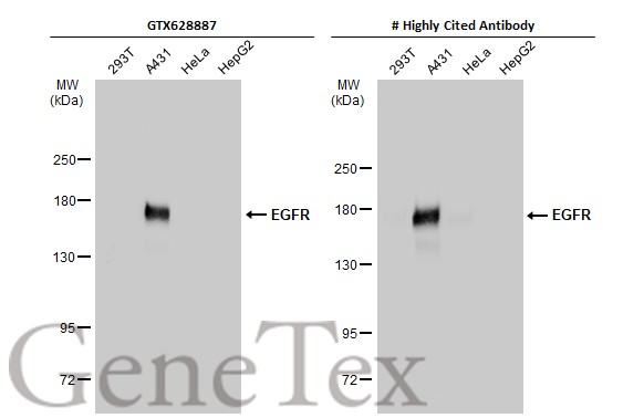
Various whole cell extracts (30 μg) were separated by 5% SDS-PAGE, and the membranes were blotted with EGFR antibody [GT133] (GTX628887) diluted at 1:5000 and competitor's antibody diluted at 1:5000. The HRP-conjugated anti-mouse IgG antibody (GTX213111-01) was used to detect the primary antibody. Corresponding RNA expression data for the same cell lines are based on Human Protein Atlas program. *The competitor is not affiliated with GeneTex and does not endorse this product. |
|
|
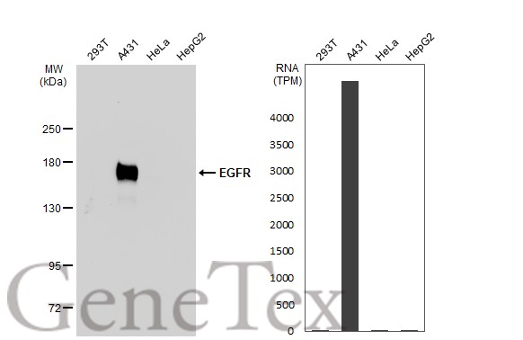
Various whole cell extracts (30 μg) were separated by 5% SDS-PAGE, and the membrane was blotted with EGFR antibody [GT133] (GTX628887) diluted at 1:5000. The HRP-conjugated anti-mouse IgG antibody (GTX213111-01) was used to detect the primary antibody. Corresponding RNA expression data for the same cell lines are based on Human Protein Atlas program. |
|
|
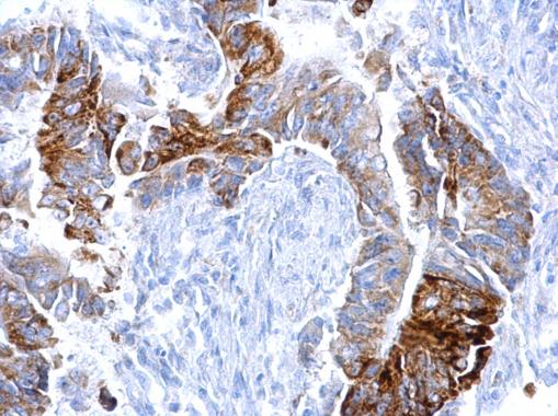
EGFR antibody [GT133] detects EGFR protein at cell membrane on human ovarian carcinoma by immunohistochemical analysis._x000D_ Sample: Paraffin-embedded human ovarian carcinoma._x000D_ EGFR antibody [GT133] (GTX628887) dilution: 1:500._x000D__x000D_ Antigen Retrieval: Trilogy™ (EDTA based, pH 8.0) buffer, 15min |
|
|
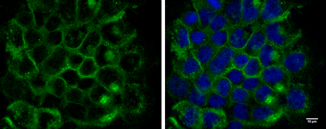
EGFR antibody [GT133] detects EGFR protein at cell membrane by immunofluorescent analysis. Sample: A431 cells were fixed in 4% paraformaldehyde for 10 min. Green: EGFR protein stained by EGFR antibody [GT133] (GTX628887) diluted at 1:300. Blue: Hoechst 33342 staining. Scale bar = 10 μm. |
|
|
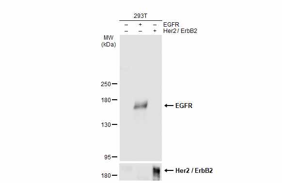
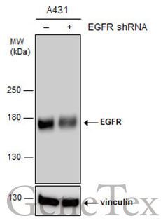
Non-transfected (–) and transfected (+) A431 whole cell extracts (15 μg) were separated by 5% SDS-PAGE, and the membrane was blotted with EGFR antibody [GT133] (GTX628887) diluted at 1:8000. The HRP-conjugated anti-mouse IgG antibody (GTX213111-01) was used to detect the primary antibody. |
|
|
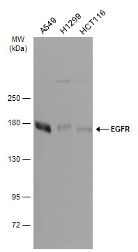
Various whole cell extracts (30 μg) were separated by 5% SDS-PAGE, and the membrane was blotted with EGFR antibody [GT133] (GTX628887) diluted at 1:10000. The HRP-conjugated anti-mouse IgG antibody (GTX213111-01) was used to detect the primary antibody. |
|
|
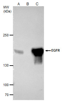
EGFR antibody [GT133] immunoprecipitates EGFR protein in IP experiments. IP samples: A431 whole cell extract A. 40 μg A431 whole cell extract B. Control with 4 μg of preimmune Mouse IgG C. Immunoprecipitation of EGFR protein by 4 μg EGFR antibody [GT133] (GTX628887) 5 % SDS-PAGE The immunoprecipitated EGFR protein was detected by EGFR antibody [GT133] (GTX628887) diluted at 1:5000. [EasyBlot anti-mouse IgG (GTX221667-01) was used as a secondary reagent] |
|
|
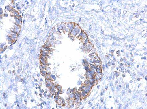
EGFR antibody [GT133] detects EGFR protein at cell membrane on human colon carcinoma by immunohistochemical analysis._x000D_ Sample: Paraffin-embedded human colon carcinoma._x000D_ EGFR antibody [GT133] (GTX628887) dilution: 1:500._x000D__x000D_ Antigen Retrieval: Trilogy™ (EDTA based, pH 8.0) buffer, 15min |
Product Guarantee and Expert Support
