
p53 antibody [DO1], IgG2a, Unconjugated, Mouse, Monoclonal
Catalog Number:
GTX70214
- Images (10)
| Article Name: | p53 antibody [DO1], IgG2a, Unconjugated, Mouse, Monoclonal |
| Biozol Catalog Number: | GTX70214 |
| Supplier Catalog Number: | GTX70214 |
| Alternative Catalog Number: | GTX70214-200 |
| Manufacturer: | GeneTex |
| Host: | Mouse |
| Category: | Antikörper |
| Application: | ELISA, FACS, ICC, IHC-Fr, IHC-P, IP, WB |
| Species Reactivity: | Human, Mouse |
| Immunogen: | Recombinant human p53 expressed in E. coli. |
| Conjugation: | Unconjugated |
| Alternative Names: | tumor protein p53 , BCC7 , BMFS5 , LFS1 , P53 , TRP53 |
| p53 antibody recognizes p53 protein, which has a predicted molecular weight of 44 kDa but is typically detected by western blot at 53 kDa. p53, known widely as a tumor suppressor, plays critical roles in various cellular processes including cell cycle re |
| Clonality: | Monoclonal |
| Concentration: | 0.25 mg/ml (Please refer to the vial label for the specific concentration.) |
| Clone Designation: | [DO1] |
| Molecular Weight: | 44 |
| Isotype: | IgG2a |
| NCBI: | 7157 |
| Pubmed: | 33061576, 27314325, |
| UniProt: | P04637 |
| Buffer: | 1XPBS (pH7), 20% Glycerol, no Preservative. |
| Source: | Human |
| Purity: | Protein A purified |
| Form: | Liquid |
| Application Notes: | WB: 1:500-1:10000. IHC-P: 1:1000. IP: 1:100-1:500. *Optimal dilutions/concentrations should be determined by the researcher.Not tested in other applications. |
|
|
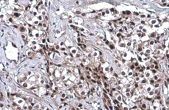
p53 antibody [DO1] detects p53 protein at nucleus by immunohistochemical analysis.Sample: Paraffin-embedded human pancreatic cancer.p53 stained by p53 antibody [DO1] (GTX70214) diluted at 1:100.Antigen Retrieval: Citrate buffer, pH 6.0, 15 min |
|
|
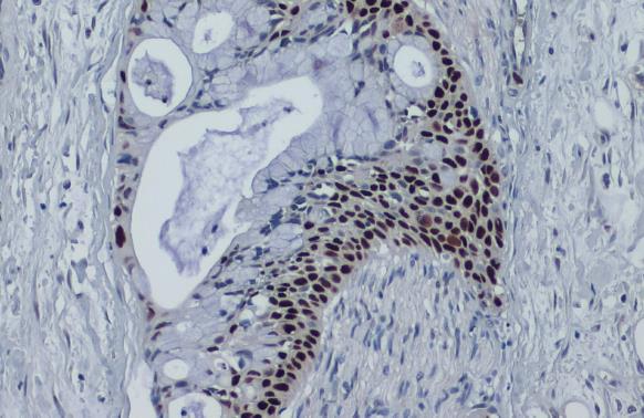
p53 antibody [DO1] detects p53 protein at cytoplasm and nucleus by immunohistochemical analysis.Sample: Paraffin-embedded human colon cancer.p53 stained by p53 antibody [DO1] (GTX70214) diluted at 1:100.Antigen Retrieval: Citrate buffer, pH 6.0, 15 min |
|
|
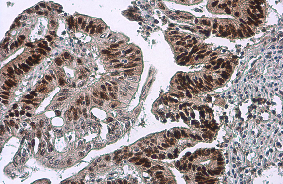
p53 antibody [DO1] detects p53 protein at cytoplasm and nucleus by immunohistochemical analysis.Sample: Paraffin-embedded human breast carcinoma.p53 stained by p53 antibody [DO1] (GTX70214) diluted at 1:100.Antigen Retrieval: Citrate buffer, pH 6.0, 15 min |
|
|
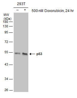
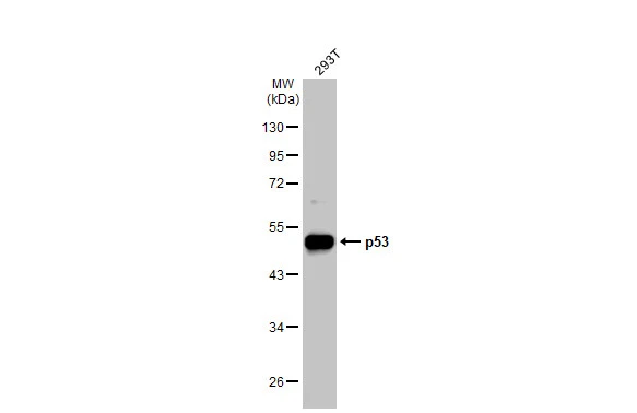
Untreated (–) and treated (+) 293T whole cell extracts (30 μg) were separated by 10% SDS-PAGE, and the membrane was blotted with p53 antibody [DO1] (GTX70214) diluted at 1:1000. The HRP-conjugated anti-mouse IgG antibody (GTX213111-01) was used to detect the primary antibody. |
|
|
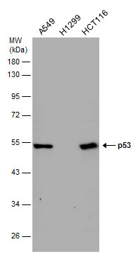
Various whole cell extracts (30 μg) were separated by 10% SDS-PAGE, and the membrane was blotted with p53 antibody [DO1] (GTX70214) diluted at 1:1000. The HRP-conjugated anti-mouse IgG antibody (GTX213111-01) was used to detect the primary antibody. |
|
|
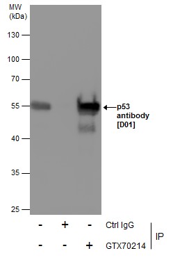
Immunoprecipitation of p53 protein from 293T whole cell extracts using 5 μg of p53 antibody [DO1] (GTX70214). Western blot analysis was performed using p53 antibody [D01] (GTX70214). EasyBlot anti-Mouse IgG (GTX221667-01) was used as a secondary reagent. |
|
|
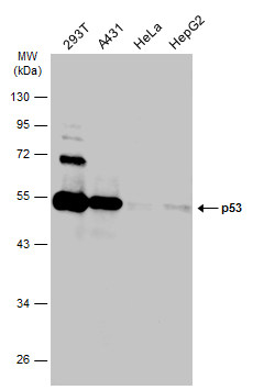
Various whole cell extracts (30 μg) were separated by 10% SDS-PAGE, and the membrane was blotted with p53 antibody [DO1] (GTX70214) diluted at 1:10000. The HRP-conjugated anti-mouset IgG antibody (GTX213111-01) was used to detect the primary antibody. |
|
|
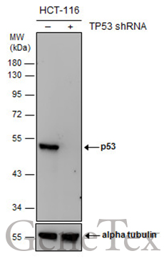
Non-transfected (–) and transfected (+) HCT116 whole cell extracts (30 μg) were separated by 10% SDS-PAGE, and the membrane was blotted with p53 antibody [DO1] (GTX70214) diluted at 1:1000. The HRP-conjugated anti-mouse IgG antibody (GTX213111-01) was used to detect the primary antibody. |
|
|
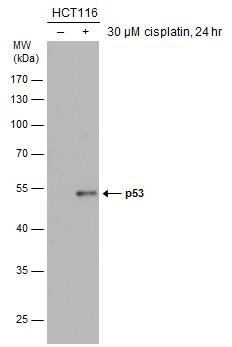
Untreated (–) and treated (+) HCT116 whole cell extracts (30 μg) were separated by 10% SDS-PAGE, and the membrane was blotted with p53 antibody [DO1] (GTX70214) diluted at 1:1000. The HRP-conjugated anti-mouse IgG antibody (GTX213111-01) was used to detect the primary antibody. |
|
|
GTX70214 WB Image |
Product Guarantee and Expert Support
