E.coli-derived human ERAB recombinant protein (Position: E48-P261). Human ERAB shares 87% and 88% amino acid (aa) sequence identity with mouse and rat ERAB, respectively.
Boster Bio Anti-ERAB/HSD17B10 Antibody Picoband catalog PB9336. Tested in Flow Cytometry, IF, IHC, ICC, WB applications. This antibody reacts with Human. The brand Picoband indicates this is a premium antibody that guarantees superior quality, high affinity, and strong signals with minimal background in Western blot applications. Only our best-performing antibodies are designated as Picoband, ensuring unmatched performance.
Klonalität:
Polyclonal
Konzentration:
Adding 0.2 ml of distilled water will yield a concentration of 500 µg/ml.
Each vial contains antibody formulated with stabilizing components, 0.9 mg NaCl, 0.2 mg Na2HPO4, and 0.05 mg NaN3. *This antibody is supplied in a stabilized formulation. Compatibility with conjugation reactions depends on the chemistry of the conjugatio
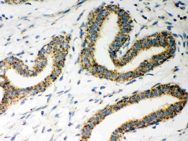
IHC analysis of ERAB using anti-ERAB antibody (PB9336).ERAB was detected in paraffin-embedded section of Human Mammary Cancer Tissue. Heat mediated antigen retrieval was performed in citrate buffer (pH6&44, epitope retrieval solution) for 20 mins. The tissue section was blocked with 10% goat serum. The tissue section was then incubated with 1µg/ml rabbit anti-ERAB Antibody (PB9336) overnight at 4C. Biotinylated goat anti-rabbit IgG was used as secondary antibody and incubated for 30 minutes at 37C. The tissue section was developed using Strepavidin-Biotin-Complex (SABC)(Catalog SA1022) with DAB as the chromogen.
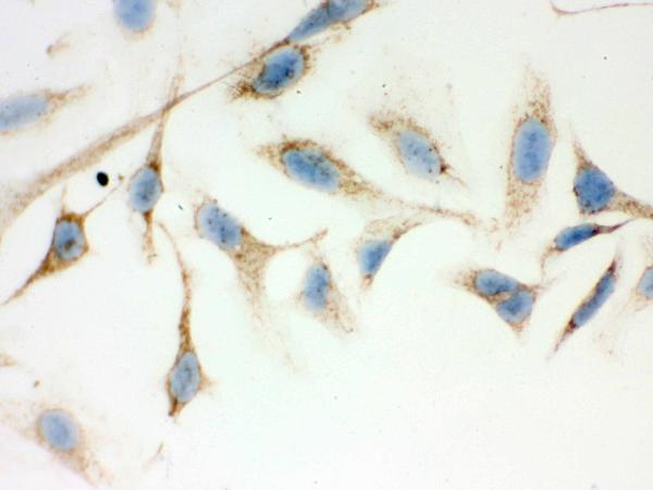
IHC analysis of ERAB using anti-ERAB antibody (PB9336). ERAB was detected in immunocytochemical section of Hela cell. Enzyme antigen retrieval was performed using IHC enzyme antigen retrieval reagent (AR0022) for 15 mins. The cells were blocked with 10% goat serum. And then incubated with 1µg/ml rabbit anti-ERAB Antibody (PB9336) overnight at 4C. Biotinylated goat anti-rabbit IgG was used as secondary antibody and incubated for 30 minutes at 37C. The section was developed using Strepavidin-Biotin-Complex (SABC)(Catalog SA1022) with DAB as the chromogen.
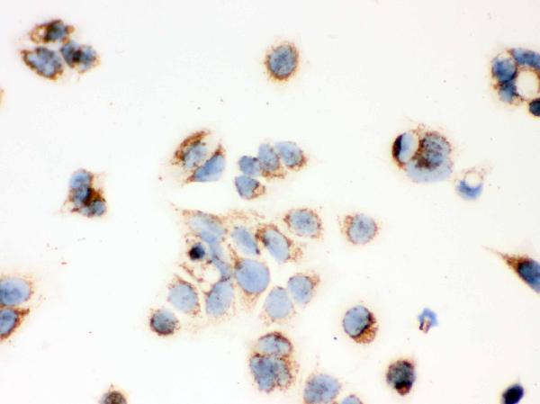
IHC analysis of ERAB using anti-ERAB antibody (PB9336). ERAB was detected in immunocytochemical section of MCF-7 cell. Enzyme antigen retrieval was performed using IHC enzyme antigen retrieval reagent (AR0022) for 15 mins. The cells were blocked with 10% goat serum. And then incubated with 1µg/ml rabbit anti-ERAB Antibody (PB9336) overnight at 4C. Biotinylated goat anti-rabbit IgG was used as secondary antibody and incubated for 30 minutes at 37C. The section was developed using Strepavidin-Biotin-Complex (SABC)(Catalog SA1022) with DAB as the chromogen.
IHC analysis of ERAB using anti-ERAB antibody (PB9336). ERAB was detected in immunocytochemical section of MCF-7 cell. Enzyme antigen retrieval was performed using IHC enzyme antigen retrieval reagent (AR0022) for 15 mins. The cells were blocked with 10% goat serum. And then incubated with 1µg/ml rabbit anti-ERAB Antibody (PB9336) overnight at 4C. Biotinylated goat anti-rabbit IgG was used as secondary antibody and incubated for 30 minutes at 37C. The section was developed using Strepavidin-Biotin-Complex (SABC)(Catalog SA1022) with DAB as the chromogen.
IHC analysis of ERAB using anti-ERAB antibody (PB9336). ERAB was detected in immunocytochemical section of SMMC-7721 cell. Enzyme antigen retrieval was performed using IHC enzyme antigen retrieval reagent (AR0022) for 15 mins. The cells were blocked with 10% goat serum. And then incubated with 1µg/ml rabbit anti-ERAB Antibody (PB9336) overnight at 4C. Biotinylated goat anti-rabbit IgG was used as secondary antibody and incubated for 30 minutes at 37C. The section was deve
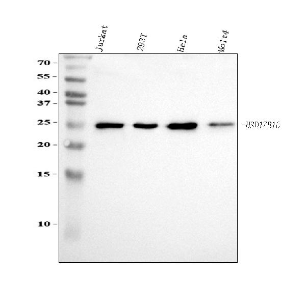
Western blot analysis of ERAB using anti-ERAB antibody (PB9336). Electrophoresis was performed on a 5-20% SDS-PAGE gel at 70V (Stacking gel) / 90V (Resolving gel) for 2-3 hours. The sample well of each lane was loaded with 30 ug of sample under reducing conditions. Lane 1: human Jurkat whole cell lysates, Lane 2: human 293T whole cell lysates, Lane 3: human Hela whole cell lysates, Lane 4: human MOLT4 whole cell lysates. After electrophoresis, proteins were transferred to a nitrocellulose membrane at 150 mA for 50-90 minutes. Blocked the membrane with 5% non-fat milk/TBS for 1.5 hour at RT. The membrane was incubated with rabbit anti-ERAB antigen affinity purified polyclonal antibody (Catalog PB9336) at 0.5 µg/mL overnight at 4C, then washed with TBS-0.1%Tween 3 times with 5 minutes each and probed with a goat anti-rabbit IgG-HRP secondary antibody at a dilution of 1:5000 for 1.5 hour at RT. The signal is developed using an Enhanced Chemiluminescent detection (ECL) kit (Catalog EK1002) with Tanon 5200 system. A specific band was detected for ERAB at approximately 25 kDa. The expected band size for ERAB is at 27-30 kDa.
* Mehrwertsteuer und Versandkosten nicht enthalten. Irrtümer und Preisänderungen vorbehalten