
AKT1 Mouse Polyclonal Antibody, Unconjugated
Artikelnummer:
BYT-ORB11276
- Bilder (9)


| Artikelname: | AKT1 Mouse Polyclonal Antibody, Unconjugated |
| Artikelnummer: | BYT-ORB11276 |
| Hersteller Artikelnummer: | orb11276 |
| Alternativnummer: | BYT-ORB11276-100,BYT-ORB11276-200,BYT-ORB11276-50 |
| Hersteller: | Biorbyt |
| Wirt: | Mouse |
| Kategorie: | Antikörper |
| Applikation: | ICC, IF, IHC-Fr, IHC-P, WB |
| Spezies Reaktivität: | Human, Mouse, Rat |
| Immunogen: | KLH conjugated synthetic peptide derived from human AKT-1 (401-479/479aa) |
| Konjugation: | Unconjugated |
| Alternative Synonym: | AKT 1, AKT, AKT-1, AKT1_HUMAN, C AKT, cAKT, MGC9965, MGC99656, Oncogene AKT1, PKB, PKB alpha, PKB-ALPHA, PRKBA, Protein Kinase B alpha, Protein kinase B, Proto-oncogene c-Akt, RAC alpha, RAC alpha serine/threonine protein kinase, RAC, RAC PK alpha, Rac protein kinase alpha, RAC Serine/Threonine Protein Kinase, RAC-alpha serine/threonine-protein kinase, RAC-PK-alpha, v akt murine thymoma viral oncogene homolog 1, vAKT Murine Thymoma Viral Oncogene Homolog 1. |
| AKT1 Mouse Polyclonal Antibody |
| Klonalität: | Polyclonal |
| Konzentration: | 1mg/ml |
| Molekulargewicht: | 56 kDa |
| UniProt: | P31749 |
| Puffer: | 0.01M TBS (pH7.4) with 1% rAlbumin, 0.02% Proclin300 and 50% Glycerol. |
| Formulierung: | Liquid |
| Target-Kategorie: | AKT1 |
| Application Verdünnung: | WB=1:500-2000, IHC-P=1:100-500, IHC-F=1:100-500, ICC/IF=1:100-500, IF=1:100-500 |
|
|
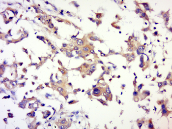
Paraformaldehyde-fixed, paraffin embedded (human memmery cancer), Antigen retrieval by boiling in sodium citrate buffer (pH6.0) for 15 min, Block endogenous peroxidase by 3% hydrogen peroxide for 20 minutes, Blocking buffer (normal goat serum) at 37C for 30 min, Antibody incubation with (AKT1) Polyclonal Antibody, Unconjugated at 1:500 overnight at 4C, followed by a conjugated secondary for 20 minutes and DAB staining. |
|
|
Sample: NIH/3T3 (Mouse) Cell Lysate at 30 ug, MCF-7 (Human) Cell Lysate at 30 ug, A549 (Human) Cell Lysate at 30 ug, Primary: Anti-AKT1 (orb11276) at 1/1000 dilution, Secondary: IRDye800CW Goat Anti-Mouse IgG at 1/20000 dilution, Predicted band size: 56 kD, Observed band size: 60 kD. |
|
|
Tissue/Cell: Hela cell, 4% Paraformaldehyde-fixed, Triton X-100 at room temperature for 20 min, Blocking buffer (normal goat serum) at 37C for 20 min, Antibody incubation with (MAKT1) polyclonal Antibody, Unconjugated (orb11276) 1:100, 90 minutes at 37C, followed by a conjugated Goat Anti-Mouse IgG-CY3 antibody at 37C for 90 minutes, DAPI (blue) was used to stain the cell nuclei. |
|
|
Tissue/Cell: MCF7 cell, 4% Paraformaldehyde-fixed, Triton X-100 at room temperature for 20 min, Blocking buffer (normal goat serum) at 37C for 20 min, Antibody incubation with (AKT1) polyclonal Antibody, Unconjugated (orb11276) 1:100, 90 minutes at 37C, followed by a CY3 conjugated Goat Anti-Mouse IgG antibody at 37C for 90 minutes, DAPI (blue) was used to stain the cell nuclei. |
|
|
Paraformaldehyde-fixed, paraffin embedded (Mouse brain), Antigen retrieval by boiling in sodium citrate buffer (pH6.0) for 15 min, Block endogenous peroxidase by 3% hydrogen peroxide for 20 minutes, Blocking buffer (normal goat serum) at 37C for 30 min, Antibody incubation with (AKT1) Monoclonal Antibody, Unconjugated (orb11276) at 1:400 overnight at 4C, followed by operating according to SP Kit (Mouse) instructionsand DAB staining. |
|
|
Paraformaldehyde-fixed, paraffin embedded (mouse liver), Antigen retrieval by boiling in sodium citrate buffer (pH6.0) for 15 min, Block endogenous peroxidase by 3% hydrogen peroxide for 20 minutes, Blocking buffer (normal goat serum) at 37C for 30 min, Antibody incubation with (AKT1) Polyclonal Antibody, Unconjugated (orb11276) at 1:200 overnight at 4C, followed by operating according to SP Kit (Rabbit) instructionsand DAB staining. |
|
|
Paraformaldehyde-fixed, paraffin embedded (rat lung), Antigen retrieval by boiling in sodium citrate buffer (pH6.0) for 15 min, Block endogenous peroxidase by 3% hydrogen peroxide for 20 minutes, Blocking buffer (normal goat serum) at 37C for 30 min, Antibody incubation with (AKT1) Monoclonal Antibody, Unconjugated (orb11276) at 1:200 overnight at 4C, followed by operating according to SP Kit (Mouse) instructionsand DAB staining. |
Produktgarantie und fachkundiger Support
