zeta chain of T cell receptor associated protein kinase 70
Anwendungsbeschreibung:
Application Notes: Western blot, 0.25-0.5 µg/ml, Human, Mouse, Rat Immunohistochemistry(Paraffin-embedded Section), 2-5 µg/ml, Human Immunocytochemistry/Immunofluorescence, 5 µg/ml, Human Flow Cytometry (Fixed), 1-3 µg/1x106 cells, Human. Adding 0.2 ml of distilled water will yield a concentration of 500 µg/ml
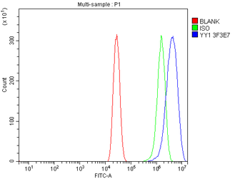
Flow Cytometry analysis of A431 cells using anti-YY1 antibody. Overlay histogram showing A431 cells (Blue line). To facilitate intracellular staining, cells were fixed with 4% paraformaldehyde and permeabilized with permeabilization buffer. The cells were blocked with 10% normal goat serum. And then incubated with mouse anti-YY1 Antibody (1 µg/1x10 6 cells) for 30 min at 20C. DyLight488 conjugated goat anti-mouse IgG (5-10 µg/1x10 6 cells) was used as secondary antibody for 30 minutes at 20C. Isotype control antibody (Green line) was mouse IgG (1 µg/1x10 6) used under the same conditions. Unlabelled sample without incubation with primary antibody and secondary antibody (Red line) was used as a blank control.
IF analysis of YY1 using anti-YY1 antibody. YY1 was detected in an immunocytochemical section of T-47D cells. Enzyme antigen retrieval was performed using IHC enzyme antigen retrieval reagent for 15 mins. The cells were blocked with 10% goat serum. And then incubated with 5 µg/mL mouse anti-YY1 Antibody overnight at 4C. DyLight594 Conjugated Goat Anti-Mouse IgG was used as secondary antibody at 1:100 dilution and incubated for 30 minutes at 37C. The section was counterstained with DAPI. Visualize using a fluorescence microscope and filter sets appropriate for the label used.
IHC analysis of YY1 using anti-YY1 antibody. YY1 was detected in a paraffin-embedded section of human laryngeal squamous cell carcinomas tissue. Heat mediated antigen retrieval was performed in EDTA buffer (pH8.0, epitope retrieval solution). The tissue section was blocked with 10% goat serum. The tissue section was then incubated with 2 µg/ml mouse anti-YY1 Antibody overnight at 4C. Peroxidase Conjugated Goat Anti-mouse IgG was used as secondary antibody and incubated for 30 minutes at 37C. The tissue section was developed using HRP Conjugated Mouse IgG Super Vision Assay Kit with DAB as the chromogen.
IHC analysis of YY1 using anti-YY1 antibody. YY1 was detected in a paraffin-embedded section of human ovarian serous cancer tissue. Heat mediated antigen retrieval was performed in EDTA buffer (pH8.0, epitope retrieval solution). The tissue section was blocked with 10% goat serum. The tissue section was then incubated with 2 µg/ml mouse anti-YY1 Antibody overnight at 4C. Peroxidase Conjugated Goat Anti-mouse IgG was used as secondary antibody and incubated for 30 minutes at 37C. The tissue section was developed using HRP Conjugated Mouse IgG Super Vision Assay Kit with DAB as the chromogen.
IHC analysis of YY1 using anti-YY1 antibody. YY1 was detected in a paraffin-embedded section of human pancreatic ductal adenocarcinoma tissue. Heat mediated antigen retrieval was performed in EDTA buffer (pH8.0, epitope retrieval solution). The tissue section was blocked with 10% goat serum. The tissue section was then incubated with 2 µg/ml mouse anti-YY1 Antibody overnight at 4C. Peroxidase Conjugated Goat Anti-mouse IgG was used as secondary antibody and incubated for 30 minutes at 37C. The tissue section was developed using HRP Conjugated Mouse IgG Super Vision Assay Kit with DAB as the chromogen.
IHC analysis of YY1 using anti-YY1 antibody. YY1 was detected in a paraffin-embedded section of human serous adenocarcinoma of ovary tissue. Heat mediated antigen retrieval was performed in EDTA buffer (pH8.0, epitope retrieval solution). The tissue section was blocked with 10% goat serum. The tissue section was then incubated with 2 µg/ml mouse anti-YY1 Antibody overnight at 4C. Peroxidase Conjugated Goat Anti-mouse IgG was used as secondary antibody and incubated for 30 minutes at 37C. The tissue section was developed using HRP Conjugated Mouse IgG Super Vision Assay Kit with DAB as the chromogen.
IHC analysis of YY1 using anti-YY1 antibody. YY1 was detected in a paraffin-embedded section of human the muscular layer of a colonic adenocarcinoma tissue. Heat mediated antigen retrieval was performed in EDTA buffer (pH8.0, epitope retrieval solution). The t
* Mehrwertsteuer und Versandkosten nicht enthalten. Irrtümer und Preisänderungen vorbehalten