
NANOG Monoclonal Antibody, IgG1, Clone: [8A1D11], Unconjugated, Mouse
Artikelnummer:
CSB-MA888008A0M
- Bilder (9)

| Artikelname: | NANOG Monoclonal Antibody, IgG1, Clone: [8A1D11], Unconjugated, Mouse |
| Artikelnummer: | CSB-MA888008A0M |
| Hersteller Artikelnummer: | CSB-MA888008A0m |
| Alternativnummer: | CSB-MA888008A0M-100UL, CSB-MA888008A0M-50UL |
| Hersteller: | Cusabio |
| Wirt: | Mouse |
| Kategorie: | Antikörper |
| Applikation: | ELISA, FC, ICC, IF, WB |
| Spezies Reaktivität: | Human, Mouse, Rat |
| Konjugation: | Unconjugated |
| Alternative Synonym: | Homeobox protein NANOG (Homeobox transcription factor Nanog) (hNanog), NANOG |
| Klonalität: | Monoclonal |
| Klon-Bezeichnung: | [8A1D11] |
| Isotyp: | IgG1 |
| UniProt: | Q9H9S0 |
| Puffer: | Preservative: 0.03% Proclin 300<br />Constituents: 50% Glycerol, 0.01M PBS, PH 7.4 |
| Reinheit: | >95%, Protein G purified |
| Formulierung: | Liquid |
| Target-Kategorie: | NANOG |
| Application Verdünnung: | Recommended dilution: WB:1:500-1:2000, ICC:1:50-1:500, IF:1:50-1:200 |
|
|
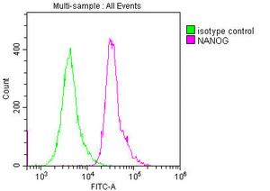
Overlay histogram showing Hela cells stained with CSB-MA888008A0m (red line) at 1:250. The cells were incubated in 1x PBS /10% normal goat serum to block non-specific protein-protein interactions followed by primary antibody for 1 h at 4C. The secondary antibody used was FITC goat anti-mouse IgG(H+L) at 1/200 dilution for 1 h at 4C. Isotype control antibody (green line) was used under the same conditions. Acquisition of >10,000 events was performed. |
|
|
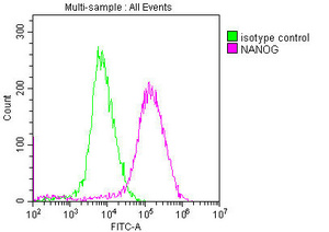
Overlay histogram showing MCF-7 cells stained with CSB-MA888008A0m (red line) at 1:250. The cells were incubated in 1x PBS /10% normal goat serum to block non-specific protein-protein interactions followed by primary antibody for 1 h at 4C. The secondary antibody used was FITC goat anti-mouse IgG(H+L) at 1/200 dilution for 1 h at 4C. Isotype control antibody (green line) was used under the same conditions. Acquisition of >10,000 events was performed. |
|
|
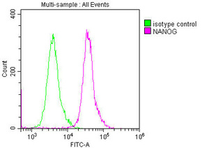
Overlay histogram showing Ntera-2 cells stained with CSB-MA888008A0m (red line) at 1:250. The cells were incubated in 1x PBS /10% normal goat serum to block non-specific protein-protein interactions followed by primary antibody for 1 h at 4C. The secondary antibody used was FITC goat anti-mouse IgG(H+L) at 1/200 dilution for 1 h at 4C. Isotype control antibody (green line) was used under the same conditions. Acquisition of >10,000 events was performed. |
|
|
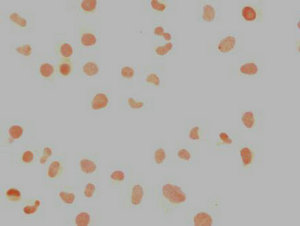
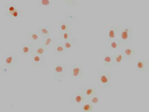
Immunocytochemistry analysis of CSB-MA888008A0m diluted at 1:100 and staining in Hela cells performed on a Leica BondTM system. The cells were fixed in 4% formaldehyde, permeabilized using 0.2% Triton X-100 and blocked with 10% normal goat serum 30min at RT. Then primary antibody (1% BSA) was incubated at 4C overnight. The primary is detected by a biotinylated secondary antibody and visualized using an HRP conjugated SP system. |
|
|
Immunocytochemistry analysis of CSB-MA888008A0m diluted at 1:100 and staining in Ntera-2 cells performed on a Leica BondTM system. The cells were fixed in 4% formaldehyde, permeabilized using 0.2% Triton X-100 and blocked with 10% normal goat serum 30min at RT. Then primary antibody (1% BSA) was incubated at 4C overnight. The primary is detected by a biotinylated secondary antibody and visualized using an HRP conjugated SP system. |
|
|
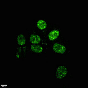
Immunofluorescence staining of Hela cells with CSB-MA888008A0m at 1:100, counter-stained with DAPI. The cells were blocked in 10% normal Goat Serum and then incubated with the primary antibody overnight at 4C. The secondary antibody was Alexa Fluor 488-congugated AffiniPure Goat Anti-Mouse IgG(H+L). |
|
|
Immunofluorescence staining of Ntera-2 cells with CSB-MA888008A0m at 1:100, counter-stained with DAPI. The cells were blocked in 10% normal Goat Serum and then incubated with the primary antibody overnight at 4C. The secondary antibody was Alexa Fluor 488-congugated AffiniPure Goat Anti-Mouse IgG(H+L). |
|
|
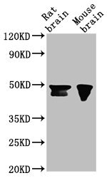
Western Blot Positive WB detected in: Rat brain tissue, Mouse brain tissue All lanes: NANOG antibody at 1:500 Secondary Goat polyclonal to Mouse IgG at 1/10000 dilution Predicted band size: 35, 33 kDa Observed band size: 46, 42 kDa |
|
|
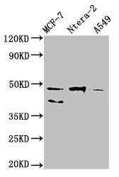
Western Blot Positive WB detected in: MCF-7 whole cell lysate, Ntera-2 whole cell lysate, A549 whole cell lysate All lanes: NANOG antibody at 1:500 Secondary Goat polyclonal to Mouse IgG at 1/10000 dilution Predicted band size: 35, 33 kDa Observed band size: 46, 40 kDa |
Produktgarantie und fachkundiger Support
