A partial length recombinant HMGB1 protein (amino acids 1- 200) was used as the immunogen for this antibody.
Alternative Names:
HMGB1||HMG1
HMGB1 (High-mobility group protein B1) is a 25 kDa non-histone chromatin protein, secreted by immune cells through leaderless secretory pathway. HMGB1 supports transcription and also interacts with nucleosomes to loosen packed DNA and remodel the chromatin. The presence of HMGB1 in the nucleus depends on posttranslational modifications. When the protein is not acetylated, it stays in the nucleus, but hyperacetylation on lysine residues causes it to translocate into the cytosol. HMGB1 has the ability to bend DNA in order to facilitate binding of other proteins to the DNA. HMGB1 has one important role in V(D)J recombination where act as a cofactor of the RAG complex. HMGB1 stimulates a plethora of cellular responses like inflammation, attracting inflammatory cells, recruiting stem cells and promoting their proliferation. HMGB1 is highly expressed in human kidney, tonsils, adipose tissue, adrenal cortex and amniotic fluid.
Western blot analysis: 2-4 µg/ml, FACS analysis: 0.5 µg/10 6 cells, Immunohistochemical analysis: 1 µg/ml
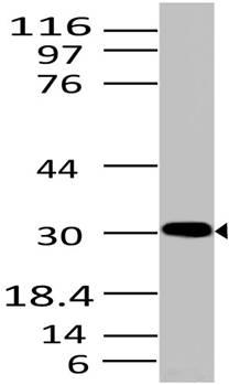
Fig-1: Western blot analysis of HMGB1. Anti- HMGB1 antibody (Clone: ABM24D3) was used at 2 µg/ml on HepG2 lysate.
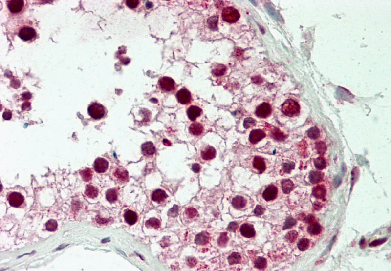
Fig-4 : Immunohistochemical analysis of HMGB1in human Testis tissue using HMGB1 antibody (Clone: ABM24D3) at 5 µg/ml.
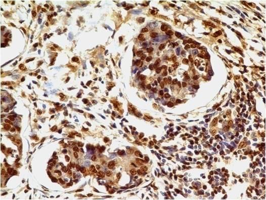
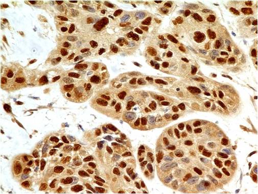
Fig-2 : Immunohistochemical analysis of HMGB1 in adenocarcinoma of stomach using HMGB1 antibody (Clone: ABM24D3) at 1 µg/ml.
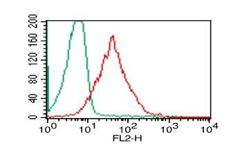
Fig-3: Intracellular flow analysis of HMGB1 in PBMC (Lymphocyte) using 0.5 µg/10 6 cells of HMGB1 antibody (Clone: ABM24D3). Green represents isotype control, red represents anti-HMGB1 antibody. Goat anti-mouse PE conjugate was used as secondary.
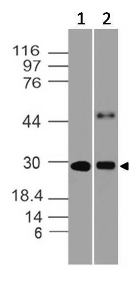
Fig-9: Western blot analysis of HMGB1. Anti- HMGB1 antibody (Clone: ABM24D3) was used at 0.5 µg/ml on (1) Ramos and (2) Raji lysates.
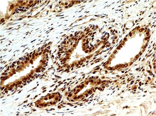
Fig-6 : Immunohistochemical analysis of HMGB1 in human Prostate using HMGB1 antibody (Clone: ABM24D3) at 1 µg/ml.
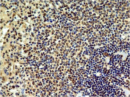
Fig-7: Immunohistochemical analysis of HMGB1in human Spleen using HMGB1 antibody (Clone: ABM24D3) at 1 µg/ml.
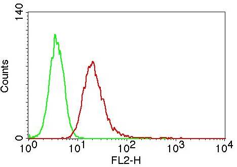
Fig-5: Intracellular flow analysis of HMGB1 in Jurkat cells using 0.5 µg/10 6 cells of HMGB1 antibody (Clone: ABM24D3). Green represents isotype control, red represents anti-HMGB1 antibody. Goat anti-mouse PE conjugate was used as secondary antibody.
Fig-8: Immunohistochemical analysis of HMGB1in squamous cell carcinoma of esophagus using HMGB1 antibody (Clone: ABM24D3) at 1 µg/ml.
* VAT and and shipping costs not included. Errors and price changes excepted