
Monoclonal Antibody to MBD2/MBD3 (Clone: ABM14A8)
Catalog Number:
ABI-10-7006
- Images (8)
| Article Name: | Monoclonal Antibody to MBD2/MBD3 (Clone: ABM14A8) |
| Biozol Catalog Number: | ABI-10-7006 |
| Supplier Catalog Number: | 10-7006 |
| Alternative Catalog Number: | ABI-10-7006-100UG |
| Manufacturer: | Abeomics |
| Host: | Mouse |
| Category: | Antikörper |
| Application: | FACS, IHC, WB |
| Species Reactivity: | Human |
| Immunogen: | A partial length recombinant MBD3 protein (amino acids 1-290) was used as the immunogen for this antibody. |
| Alternative Names: | MBD3 |
| MBD2 is a subunit of the NuRD complex that is postulated to mediate gene repression via recruitment of the complex to methylated DNA. This protein binds around 1 kb downstream of the transcription start site of a subset of 400 CpG island promoters that are characterized by the presence of active histone marks, RNA polymerase II (Pol2) and low to medium gene expression levels and H3K36me3 deposition. Methylated MBD2 is involved in silencing methylated tumor suppressor genes as well as activation of pro-metastatic genes. MBD2 also plays a key role in regulating expression of a range of genes that are associated with optimal DC (Dendritic cells) activation and function. |
| Application Dilute: | Western blot analysis: 2-4 µg/ml, Immunohistochemical analysis: 5 µg/ml, FACS analysis: 0.5 µg/10 6 cells |
|
|
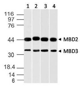
Fig-1: Western blot analysis of MBD3 (and MBD2). Anti-MBD3 (and MBD2) antibody (Clone: ABM14A8) was used at 2 µg/ml on A431, K562, A375 and HEK293 lysates. |
|
|
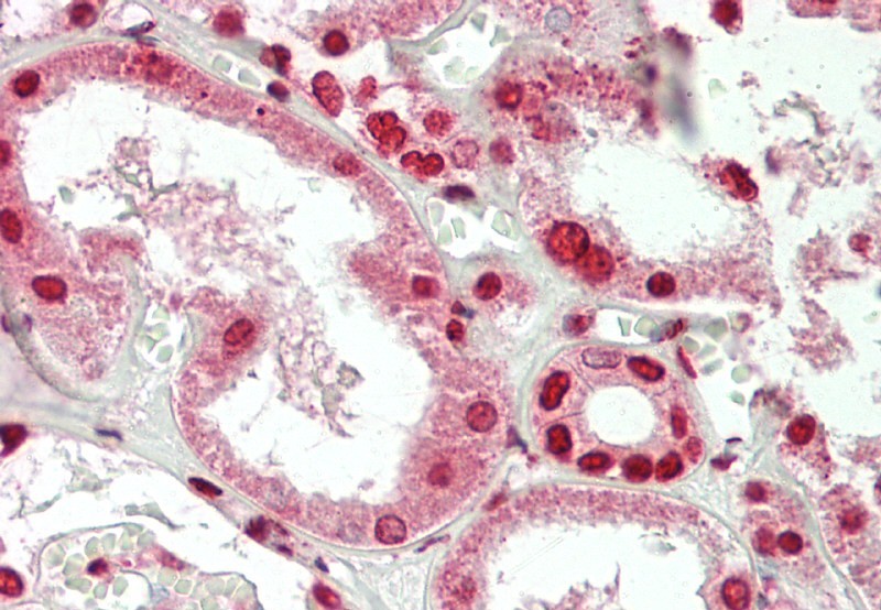
Fig-3 : Immunohistochemical analysis of MBD3 in human Kidney tissue using MBD3 antibody (Clone: ABM14A8) at 10 µg/ml. |
|
|
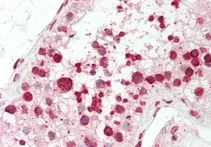
Fig-4 : Immunohistochemical analysis of MBD3 in human Testis tissue using MBD3 antibody (Clone: ABM14A8) at 10 µg/ml. |
|
|
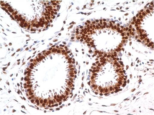
Fig-2 : Immunohistochemical analysis of MBD3 in human breast tissue using MBD3 antibody (Clone: ABM14A8) at 5 µg/ml. |
|
|
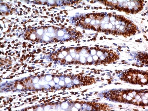
Fig-6 : Immunohistochemical analysis of MBD3 in human Colon tissue using MBD3 antibody (Clone: ABM14A8) at 5 µg/ml. |
|
|
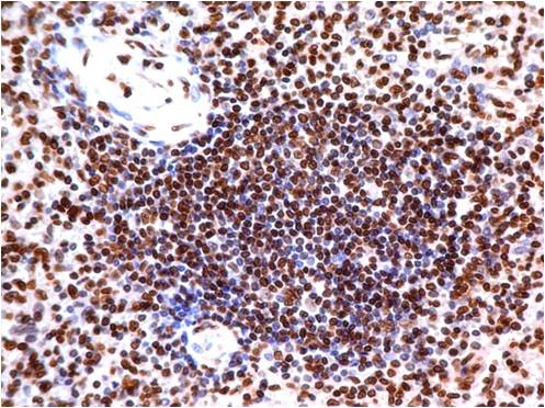
Fig-7 : Immunohistochemical analysis of MBD3 in human Spleen tissue using MBD3 antibody (Clone: ABM14A8) at 5 µg/ml. |
|
|
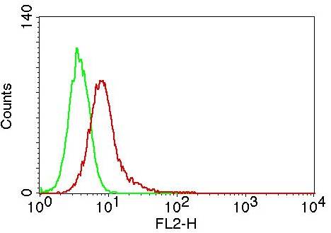
Fig-5: Intracellular flow analysis of MBD2/MBD3 in Jurkat cells using 0.5 µg/10 6 cells of MBD2/MBD3 antibody (Clone: ABM14A8). Green represents isotype control, red represents anti-MBD2/MBD3 antibody. Goat anti-mouse PE conjugate was used as secondary antibody. |
|
|
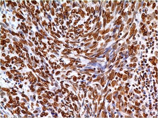
Fig-8 : Immunohistochemical analysis of MBD3 in Squamous cell carcinoma of Lungs using MBD3 antibody (Clone: ABM14A8) at 5 µg/ml. |
Product Guarantee and Expert Support
