Application Notes: Western blot, 0.25-0.5 µg/ml, Human Immunohistochemistry(Paraffin-embedded Section), 2-5 µg/ml, Human, Mouse, Rat. Adding 0.2 ml of distilled water will yield a concentration of 500 µg/ml
IHC analysis of PC4/SUB1 using anti-PC4/SUB1 antibody. PC4/SUB1 was detected in a paraffin-embedded section of human colonic adenoma tissue. Heat mediated antigen retrieval was performed in EDTA buffer (pH8.0, epitope retrieval solution). The tissue section was blocked with 10% goat serum. The tissue section was then incubated with 2 µg/ml mouse anti-PC4/SUB1 Antibody overnight at 4C. Peroxidase Conjugated Goat Anti-mouse IgG was used as secondary antibody and incubated for 30 minutes at 37C. The tissue section was developed using HRP Conjugated Mouse IgG Super Vision Assay Kit with DAB as the chromogen.
IHC analysis of PC4/SUB1 using anti-PC4/SUB1 antibody. PC4/SUB1 was detected in a paraffin-embedded section of human laryngeal squamous cell carcinoma tissue. Heat mediated antigen retrieval was performed in EDTA buffer (pH8.0, epitope retrieval solution). The tissue section was blocked with 10% goat serum. The tissue section was then incubated with 2 µg/ml mouse anti-PC4/SUB1 Antibody overnight at 4C. Peroxidase Conjugated Goat Anti-mouse IgG was used as secondary antibody and incubated for 30 minutes at 37C. The tissue section was developed using HRP Conjugated Mouse IgG Super Vision Assay Kit with DAB as the chromogen.
IHC analysis of PC4/SUB1 using anti-PC4/SUB1 antibody. PC4/SUB1 was detected in a paraffin-embedded section of human liver cancer tissue. Heat mediated antigen retrieval was performed in EDTA buffer (pH8.0, epitope retrieval solution). The tissue section was blocked with 10% goat serum. The tissue section was then incubated with 2 µg/ml mouse anti-PC4/SUB1 Antibody overnight at 4C. Peroxidase Conjugated Goat Anti-mouse IgG was used as secondary antibody and incubated for 30 minutes at 37C. The tissue section was developed using HRP Conjugated Mouse IgG Super Vision Assay Kit with DAB as the chromogen.
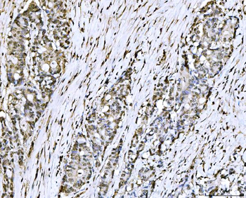
IHC analysis of PC4/SUB1 using anti-PC4/SUB1 antibody. PC4/SUB1 was detected in a paraffin-embedded section of human lung cancer tissue. Heat mediated antigen retrieval was performed in EDTA buffer (pH8.0, epitope retrieval solution). The tissue section was blocked with 10% goat serum. The tissue section was then incubated with 2 µg/ml mouse anti-PC4/SUB1 Antibody overnight at 4C. Peroxidase Conjugated Goat Anti-mouse IgG was used as secondary antibody and incubated for 30 minutes at 37C. The tissue section was developed using HRP Conjugated Mouse IgG Super Vision Assay Kit with DAB as the chromogen.
IHC analysis of PC4/SUB1 using anti-PC4/SUB1 antibody. PC4/SUB1 was detected in a paraffin-embedded section of human poison impregnated thyroid gland tissue. Heat mediated antigen retrieval was performed in EDTA buffer (pH8.0, epitope retrieval solution). The tissue section was blocked with 10% goat serum. The tissue section was then incubated with 2 µg/ml mouse anti-PC4/SUB1 Antibody overnight at 4C. Peroxidase Conjugated Goat Anti-mouse IgG was used as secondary antibody and incubated for 30 minutes at 37C. The tissue section was developed using HRP Conjugated Mouse IgG Super Vision Assay Kit with DAB as the chromogen.
IHC analysis of PC4/SUB1 using anti-PC4/SUB1 antibody. PC4/SUB1 was detected in a paraffin-embedded section of human renal cell carcinoma tissue. Heat mediated antigen retrieval was performed in EDTA buffer (pH8.0, epitope retrieval solution). The tissue section was blocked with 10% goat serum. The tissue section was then incubated with 2 µg/ml mouse anti-PC4/SUB1 Antibody overnight at 4C. Peroxidase Conjugated Goat Anti-mouse IgG was used as secondary antibody and incubated for 30 minutes at 37C. The tissue section was developed using HRP Conjugated Mouse IgG Super Vision Assay Kit with DAB as the chromogen.
IHC analysis of PC4/SUB1 using anti-PC4/SUB1 antibody. PC4/SUB1 was detected in a paraffin-embedded section of mouse colon tissue. Heat mediated antigen retrieval was performed in EDTA buffer (pH8.0, epitope retrieval solution). The tissue section was blocked with 10% goat serum. T
* VAT and and shipping costs not included. Errors and price changes excepted