
ENO1 Monoclonal Antibody, IgG1, Clone: [4D11F5], Unconjugated, Mouse
Catalog Number:
CSB-MA007670A1M
- Images (9)
| Article Name: | ENO1 Monoclonal Antibody, IgG1, Clone: [4D11F5], Unconjugated, Mouse |
| Biozol Catalog Number: | CSB-MA007670A1M |
| Supplier Catalog Number: | CSB-MA007670A1m |
| Alternative Catalog Number: | CSB-MA007670A1M-100UL, CSB-MA007670A1M-50UL |
| Manufacturer: | Cusabio |
| Host: | Mouse |
| Category: | Antikörper |
| Application: | ELISA, FC, IF, IP, WB |
| Species Reactivity: | Human, Mouse, Rabbit, Rat |
| Conjugation: | Unconjugated |
| Alternative Names: | Alpha-enolase (2-phospho-D-glycerate hydro-lyase) (C-myc promoter-binding protein) (Enolase 1) (MBP-1) (MPB-1) (Non-neural enolase) ( NNE) (Phosphopyruvate hydratase) (Plasminogen-binding protein), ENO1, ENO1L1, MBPB1, MPB1 |
| Clonality: | Monoclonal |
| Clone Designation: | [4D11F5] |
| Isotype: | IgG1 |
| UniProt: | P06733 |
| Buffer: | Preservative: 0.03% Proclin 300<br />Constituents: 50% Glycerol, 0.01M PBS, PH 7.4 |
| Purity: | >95%, Protein G purified |
| Form: | Liquid |
| Target: | ENO1 |
| Application Dilute: | Recommended dilution: WB: 1:5000-1:640000, IF:1:50-1:300, FC:1:100-1:600, IP:1µl-4µl |
|
|
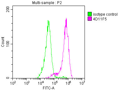
Overlay histogram showing MCF-7 cells stained with CSB-MA007670A1m (red line) at 1:550. The cells were incubated in 1x PBS /10% normal goat serum to block non-specific protein-protein interactions followed by primary antibody for 1 h at 4C. The secondary antibody used was FITC goat anti-mouse IgG(H+L) at 1/200 dilution for 1 h at 4C. Isotype control antibody (green line) was used under the same conditions. Acquisition of >10,000 events was performed. |
|
|
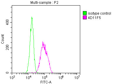
Overlay histogram showing Hela cells stained with CSB-MA007670A1m (red line) at 1:550. The cells were incubated in 1x PBS /10% normal goat serum to block non-specific protein-protein interactions followed by primary antibody for 1 h at 4C. The secondary antibody used was FITC goat anti-mouse IgG(H+L) at 1/200 dilution for 1 h at 4C. Isotype control antibody (green line) was used under the same conditions. Acquisition of >10,000 events was performed. |
|
|
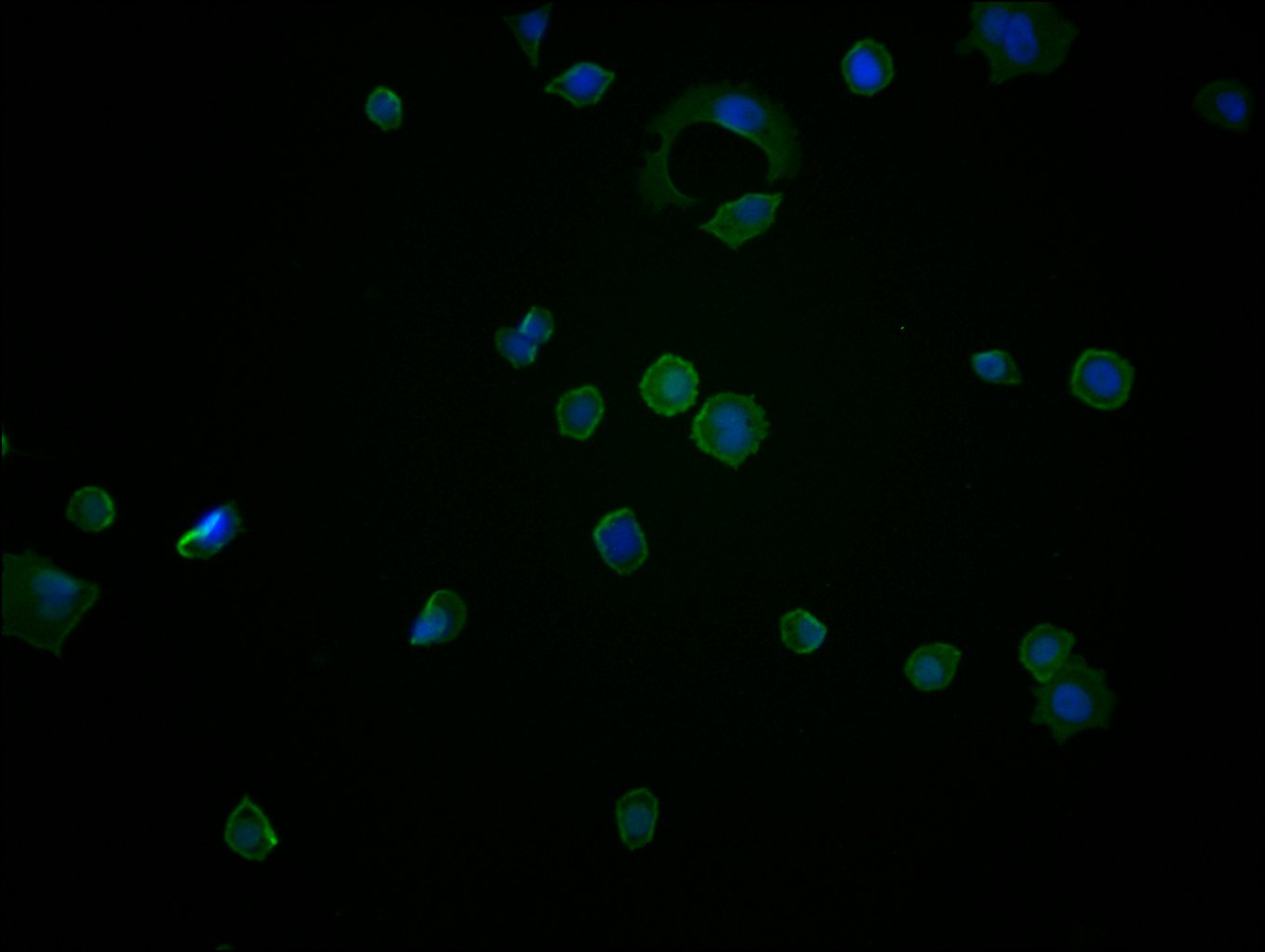
Immunofluorescence staining of MCF-7 cells with CSB-MA007670A0m at 1:270, counter-stained with DAPI. The cells were blocked in 10% normal Goat Serum and then incubated with the primary antibody overnight at 4C. The secondary antibody was Alexa Fluor 488-congugated AffiniPure Goat Anti-Mouse IgG(H+L). |
|
|
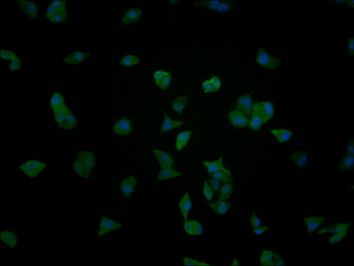
Immunofluorescence staining of Hela cells with CSB-MA007670A0m at 1:270, counter-stained with DAPI. The cells were blocked in 10% normal Goat Serum and then incubated with the primary antibody overnight at 4C. The secondary antibody was Alexa Fluor 488-congugated AffiniPure Goat Anti-Mouse IgG(H+L). |
|
|
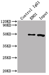
Immunoprecipitating ENO1 in HepG2 whole cell lysate Lane 1: Mouse control IgG (1µg) instead of CSB-MA007670A1m in HepG2 whole cell lysate. For western blotting, a HRP-conjugated Protein G antibody was used as the secondary antibody (1/5000) Lane 2: CSB-MA007670A1m (1µl) + HepG2 whole cell lysate (500µg) Lane 3: HepG2 whole cell lysate (10µg) |
|
|
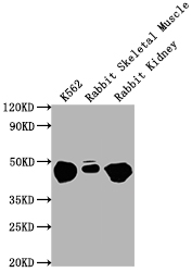
Western Blot Positive WB detected in: K562 whole cell lysate, Rabbit Skeletal Muscle tissue, Rabbit Kidney lysate All lanes ENO1 antibody at 1:10000 Secondary Goat polyclonal to mouse IgG at 0.261ug/ml Predicted band size: 47 KDa Observed band size: 47 KDa Exposure time: 1min |
|
|
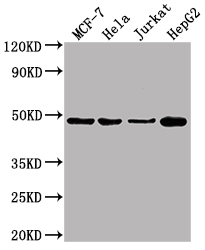
Western Blot Positive WB detected in: MCF-7 whole cell lysate, Hela whole cell lysate, Jurkat whole cell lysate, HepG2 whole cell lysate All lanes ENO1 antibody at 1:10000 Secondary Goat polyclonal to mouse IgG at 0.261ug/ml Predicted band size: 47 KDa Observed band size: 47 KDa Exposure time: 10s |
|
|
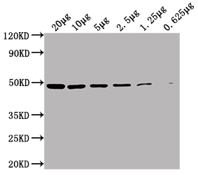
Western Blot Positive WB detected in: HepG2 whole cell lysate at 20µg, 10µg, 5µg, 2.5µg, 1.25µg, 0.625µg All lanes: ENO1 antibody at 1:5000 Secondary Goat polyclonal to Mouse IgG at 1/10000 dilution Predicted band size: 47 kDa Observed band size: 47 KDa Exposure time: 10s |
|
|
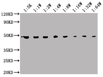
Western Blot Positive WB detected in: MCF-7 whole cell lysate All lanes: ENO1 antibody at 1:5000, 1:10000, 1:20000, 1:40000, 1:80000, 1:160000, 1:320000, 1:640000 Secondary Goat polyclonal to Mouse IgG at 1/10000 dilution Predicted band size: 47 kDa Observed band size: 47 KDa Exposure time: 10s |
Product Guarantee and Expert Support
