
GFAP Monoclonal Antibody, IgG2b, Clone: [1C91F1], Unconjugated, Mouse
Catalog Number:
CSB-MA009369A0M
- Images (9)
| Article Name: | GFAP Monoclonal Antibody, IgG2b, Clone: [1C91F1], Unconjugated, Mouse |
| Biozol Catalog Number: | CSB-MA009369A0M |
| Supplier Catalog Number: | CSB-MA009369A0m |
| Alternative Catalog Number: | CSB-MA009369A0M-100UL, CSB-MA009369A0M-50UL |
| Manufacturer: | Cusabio |
| Host: | Mouse |
| Category: | Antikörper |
| Application: | ELISA, FC, IF, IHC, WB |
| Species Reactivity: | Human, Mouse, Rat |
| Conjugation: | Unconjugated |
| Alternative Names: | Glial fibrillary acidic protein, GFAP |
| Clonality: | Monoclonal |
| Clone Designation: | [1C91F1] |
| Isotype: | IgG2b |
| UniProt: | P14136 |
| Buffer: | Preservative: 0.03% Proclin 300<br />Constituents: 50% Glycerol, 0.01M PBS, PH 7.4 |
| Purity: | >95%, Protein G purified |
| Form: | Liquid |
| Target: | GFAP |
| Application Dilute: | Recommended dilution: WB:1:500-1:5000, IHC:1:50-1:500, IF:1:50-1:200 |
|
|
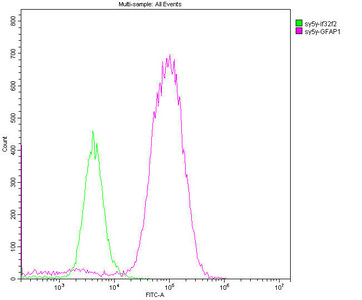
Overlay histogram showing SH-SY5Y cells stained with CSB-MA009369A0m (red line). The cells were fixed with 70% Ethylalcohol (18h) and then permeabilized with 0.3% Triton X-100 for 2 min. The cells were then incubated in 1x PBS /10% normal goat serum to block non-specific protein-protein interactions followed by the antibody (10µg/1*106cells) for 1 h at 4C. The secondary antibody used was FITC goat anti-mouse IgG(H+L) at 1/200 dilution for 1 h at 4C. Isotype control antibody (green line) was mouse IgG2b (10µg/1*106cells) used under the same conditions. Acquisition of >10,000 events was performed. |
|
|
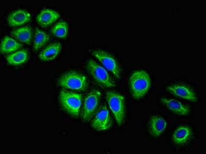
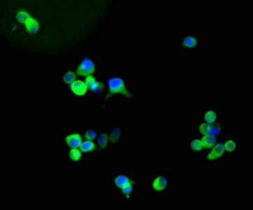
Immunofluorescent analysis of A549 cells using CSB-MA009369A0m at a dilution of 1:100 and Alexa Fluor 488-congugated AffiniPure Goat Anti-Mouse IgG(H+L). |
|
|
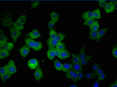
Immunofluorescent analysis of SH-SY5Y cells using CSB-MA009369A0m at a dilution of 1:100 and Alexa Fluor 488-congugated AffiniPure Goat Anti-Mouse IgG(H+L). |
|
|
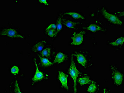
Immunofluorescent analysis of U251 cells using CSB-MA009369A0m at a dilution of 1:100 and Alexa Fluor 488-congugated AffiniPure Goat Anti-Mouse IgG(H+L). |
|
|
Immunofluorescent analysis of U87 cells using CSB-MA009369A0m at a dilution of 1:100 and Alexa Fluor 488-congugated AffiniPure Goat Anti-Mouse IgG(H+L). |
|
|
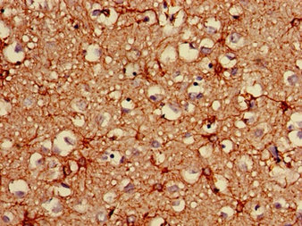
Immunohistochemistry of paraffin-embedded human brain tissue using CSB-MA009369A0m at dilution of 1:100 |
|
|
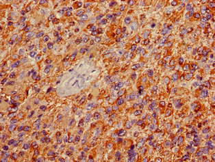
Immunohistochemistry of paraffin-embedded human glioma using CSB-MA009369A0m at dilution of 1:100 |
|
|
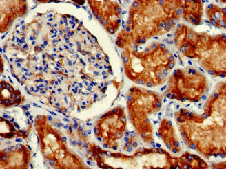
Immunohistochemistry of paraffin-embedded human kidney tissue using CSB-MA009369A0m at dilution of 1:100 |
|
|
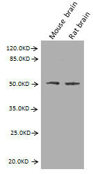
Western Blot Positive WB detected in: Mouse brain tissue, Rat brain tissue All lanes: GFAP antibody at 2.7µg/ml Secondary Goat polyclonal to Mouse IgG at 1/10000 dilution Predicted band size: 50, 51 kDa Observed band size: 50 kDa |
Product Guarantee and Expert Support
