
HSPA8 Monoclonal Antibody, IgG1, Clone: [6E4D2], Unconjugated, Mouse
Catalog Number:
CSB-MA010829A1M
- Images (9)
| Article Name: | HSPA8 Monoclonal Antibody, IgG1, Clone: [6E4D2], Unconjugated, Mouse |
| Biozol Catalog Number: | CSB-MA010829A1M |
| Supplier Catalog Number: | CSB-MA010829A1m |
| Alternative Catalog Number: | CSB-MA010829A1M-100UL, CSB-MA010829A1M-50UL |
| Manufacturer: | Cusabio |
| Host: | Mouse |
| Category: | Antikörper |
| Application: | ELISA, FC, IF, IHC, WB |
| Species Reactivity: | Human |
| Conjugation: | Unconjugated |
| Alternative Names: | Heat shock cognate 71 kDa protein (Heat shock 70 kDa protein 8) (Lipopolysaccharide-associated protein 1) (LAP-1) (LPS-associated protein 1), HSPA8, HSC70 HSP73 HSPA10 |
| Clonality: | Monoclonal |
| Clone Designation: | [6E4D2] |
| Isotype: | IgG1 |
| UniProt: | P11142 |
| Buffer: | Preservative: 0.03% Proclin 300<br />Constituents: 50% Glycerol, 0.01M PBS, PH 7.4 |
| Purity: | >95%, Protein G purified |
| Form: | Liquid |
| Target: | HSPA8 |
| Application Dilute: | Recommended dilution: WB: 1:5000-1:32000, IHC: 1:100-1:500, IF: 1:100-1:300, FC: 1:100-1:300 |
|
|
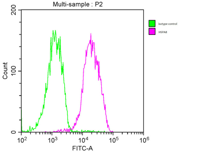
Overlay histogram showing MCF-7 cells stained with CSB-MA010829A1m (red line). The cells were fixed with 70% Ethylalcohol (18h) and then incubated in 10% normal goat serum to block non-specific protein-protein interactions followed by the primary antibody at 1/200 for 1 h at 4C. The secondary antibody used was FITC goat anti-mouse IgG(H+L) at 1/100 dilution for 30min at 4C. Isotype control antibody (green line) was mouse IgG1 used under the same conditions. Acquisition of >10,000 events was performed. |
|
|
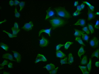
Immunofluorescence staining of Hela cells with CSB-MA010829A1m at 1:100, counter-stained with DAPI. The cells were fixed in 4% formaldehyde and blocked in 10% normal Goat Serum. The cells were then incubated with the antibody overnight at 4C. Nuclear DNA was labeled in blue with DAPI. The secondary antibody was FITC-conjugated AffiniPure Goat Anti-Mouse IgG (H+L). |
|
|
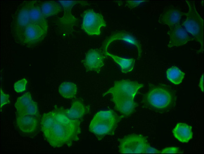
Immunofluorescence staining of MCF-7 cells with CSB-MA010829A1m at 1:100, counter-stained with DAPI. The cells were fixed in 4% formaldehyde and blocked in 10% normal Goat Serum. The cells were then incubated with the antibody overnight at 4C. Nuclear DNA was labeled in blue with DAPI. The secondary antibody was FITC-conjugated AffiniPure Goat Anti-Mouse IgG (H+L). |
|
|
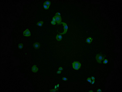
Immunofluorescence staining of PC-3 cells with CSB-MA010829A1m at 1:100, counter-stained with DAPI. The cells were fixed in 4% formaldehyde and blocked in 10% normal Goat Serum. The cells were then incubated with the antibody overnight at 4C. Nuclear DNA was labeled in blue with DAPI. The secondary antibody was FITC-conjugated AffiniPure Goat Anti-Mouse IgG (H+L). |
|
|
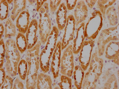
IHC image of CSB-MA010829A1m diluted at 1:220 and staining in paraffin-embedded human kidney tissue performed on a Leica BondTM system. After dewaxing and hydration, antigen retrieval was mediated by high pressure in a citrate buffer (pH 6.0). Section was blocked with 10% normal goat serum 30min at RT. Then primary antibody (1% BSA) was incubated at 4C overnight. The primary is detected by a biotinylated secondary antibody and visualized using an HRP conjugated SP system. |
|
|
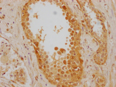
IHC image of CSB-MA010829A1m diluted at 1:220 and staining in paraffin-embedded human prostate cancer performed on a Leica BondTM system. After dewaxing and hydration, antigen retrieval was mediated by high pressure in a citrate buffer (pH 6.0). Section was blocked with 10% normal goat serum 30min at RT. Then primary antibody (1% BSA) was incubated at 4C overnight. The primary is detected by a biotinylated secondary antibody and visualized using an HRP conjugated SP system. |
|
|
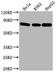
Western Blot Positive WB detected in: Hela whole cell lysate, K562 whole cell lysate, HepG2 whole cell lysate All lanes HSPA8 antibody at 1:2000 Secondary Goat polyclonal to mouse IgG at 1/50000 dilution Predicted band size: 70~75 KDa Observed band size: 70~75 KDa Exposure time: 5min |
|
|
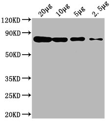
Western Blot Positive WB detected in: Hela whole cell lysate at 20ug, 10ug, 5ug, 2.5ug All lanes: HSPA8 antibody at 1:2000 Secondary Goat polyclonal to mouse IgG at 1/50000 dilution Predicted band size: 70~75 KDa Observed band size: 70~75 KDa Exposure time: 5min |
|
|
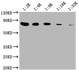
Western Blot Positive WB detected in: 20ug hela whole cell lysate HSPA8 antibody at 1:2000, 1:4000, 1:8000, 1:16000, 1:32000 Secondary Goat polyclonal to mouse IgG at 1/50000 dilution Predicted band size: 70~75 KDa Observed band size: 70~75 KDa Exposure time: 5min |
Product Guarantee and Expert Support
