
ANXA11 Antibody, Unconjugated, Rabbit, Polyclonal
Catalog Number:
CSB-PA001838ESR2HU
- Images (9)


| Article Name: | ANXA11 Antibody, Unconjugated, Rabbit, Polyclonal |
| Biozol Catalog Number: | CSB-PA001838ESR2HU |
| Supplier Catalog Number: | CSB-PA001838ESR2HU |
| Alternative Catalog Number: | CSB-PA001838ESR2HU-100UL |
| Manufacturer: | Cusabio |
| Host: | Rabbit |
| Category: | Antikörper |
| Application: | ELISA, IF, IHC, WB |
| Species Reactivity: | Human |
| Conjugation: | Unconjugated |
| Alternative Names: | 56 kDa autoantigen antibody, Annexin A11 antibody, Annexin XI antibody, Annexin-11 antibody, AnnexinA11 antibody, AnnexinXI antibody, ANX 11 antibody, ANX A11 antibody, ANX11 antibody, ANX11_HUMAN antibody, ANXA 11 antibody, ANXA11 antibody, Autoantigen 56 kD antibody, Calcyclin associated annexin 50 antibody, Calcyclin-associated annexin 50 antibody, CAP 50 antibody, CAP-50 antibody, CAP50 antibody, OTTHUMP00000059806 antibody |
| Clonality: | Polyclonal |
| UniProt: | P50995 |
| Buffer: | Preservative: 0.02% sodium azide<br />Constituents: 50% Glycerol, 0.01M PBS, pH 7.4 |
| Purity: | Antigen Affinity Purified |
| Form: | Liquid |
| Target: | ANXA11 |
| Application Dilute: | Recommended dilution: WB:1:1000-1:3000, IHC:1:50-1:200, IF:1:20-1:100 |
|
|
Immunohistochemist |
|
|
|
|
|
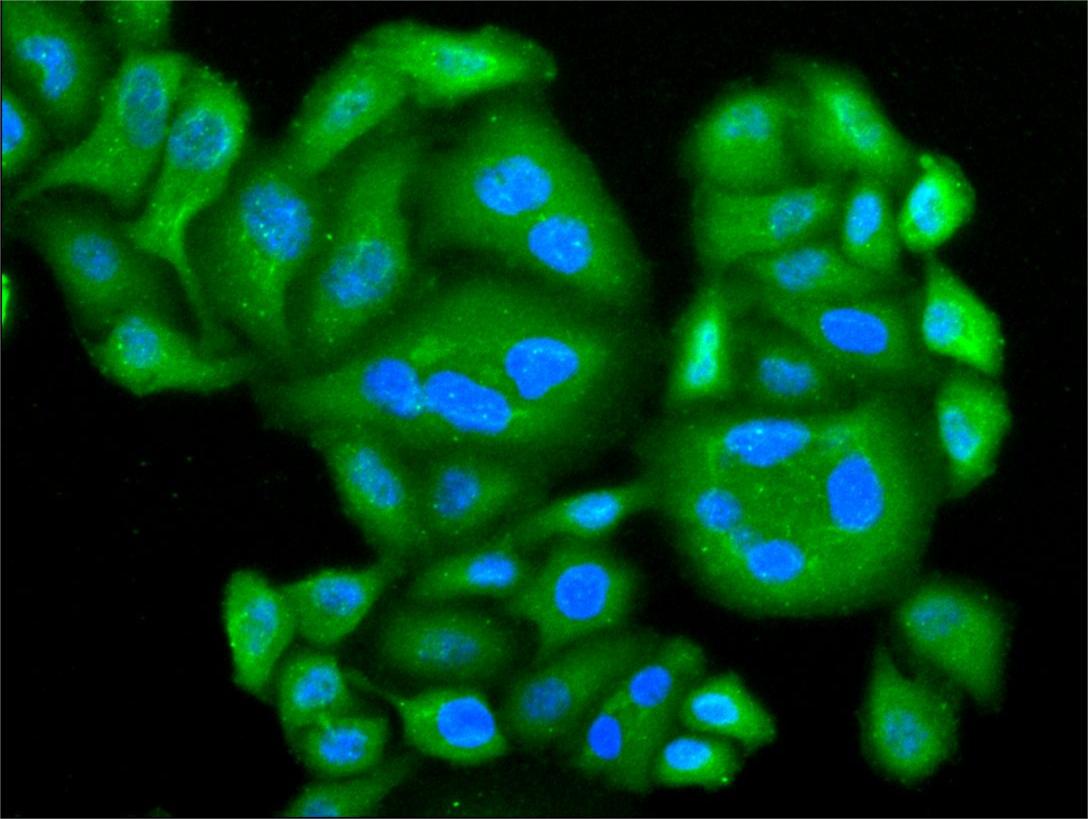
Immunofluorescence staining of A549 cell with CSB-PA001838ESR2HU at 1:20, counter-stained with DAPI. The cells were fixed in 4% formaldehyde and blocked in 10% normal Goat Serum. The cells were then incubated with the antibody overnight at 4C. The secondary antibody was Alexa Fluor 488-congugated AffiniPure Goat Anti-Rabbit IgG(H+L). |
|
|
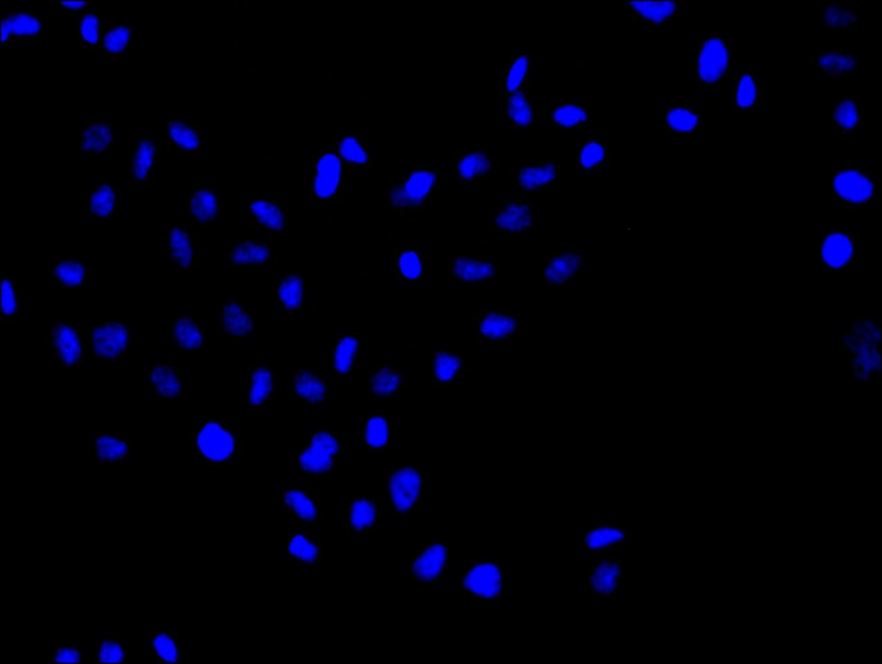
ImmunofluorescencestainingofA549cellwith5%goatserum,counter-stainedwithDAPI.Thecellswerefixedin4%formaldehydeandblockedin10%normalGoatSerum.Thecellswerethenincubatedwiththeantibodyovernightat4C.ThesecondaryantibodywasAlexaFluor488-congugatedAffiniPureGoatAnti-RabbitIgG(H+L). |
|
|
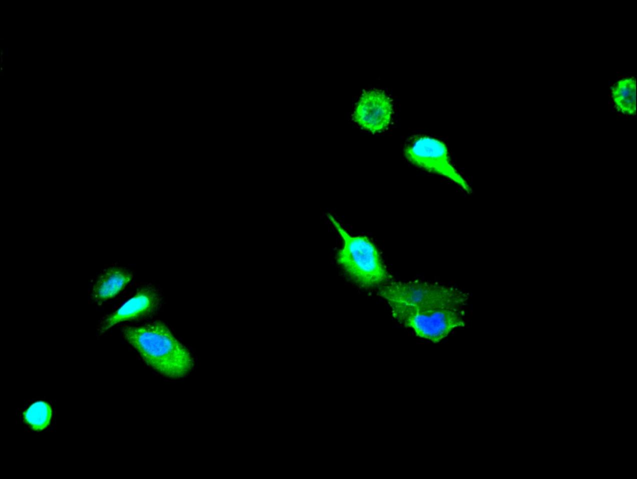
Immunofluorescence staining of Hela cell with CSB-PA001838ESR2HU at 1:20, counter-stained with DAPI. The cells were fixed in 4% formaldehyde and blocked in 10% normal Goat Serum. The cells were then incubated with the antibody overnight at 4C. The secondary antibody was Alexa Fluor 488-congugated AffiniPure Goat Anti-Rabbit IgG(H+L). |
|
|
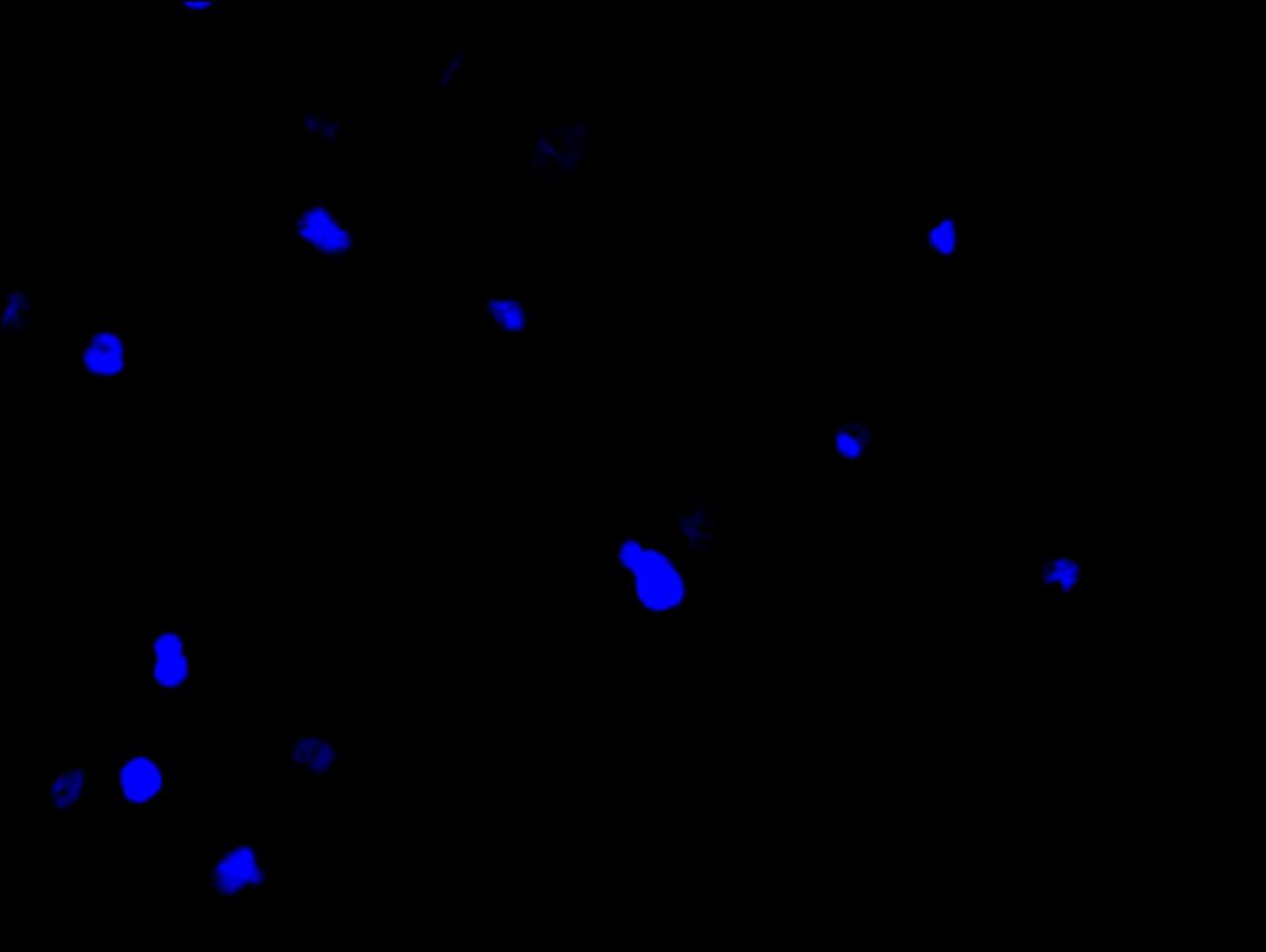
Immunofluorescence staining of Hela cell with 5% goat serum, counter-stained with DAPI. The cells were fixed in 4% formaldehyde and blocked in 10% normal Goat Serum. The cells were then incubated with the antibody overnight at 4C. The secondary antibody was Alexa Fluor 488-congugated AffiniPure Goat Anti-Rabbit IgG(H+L). |
|
|
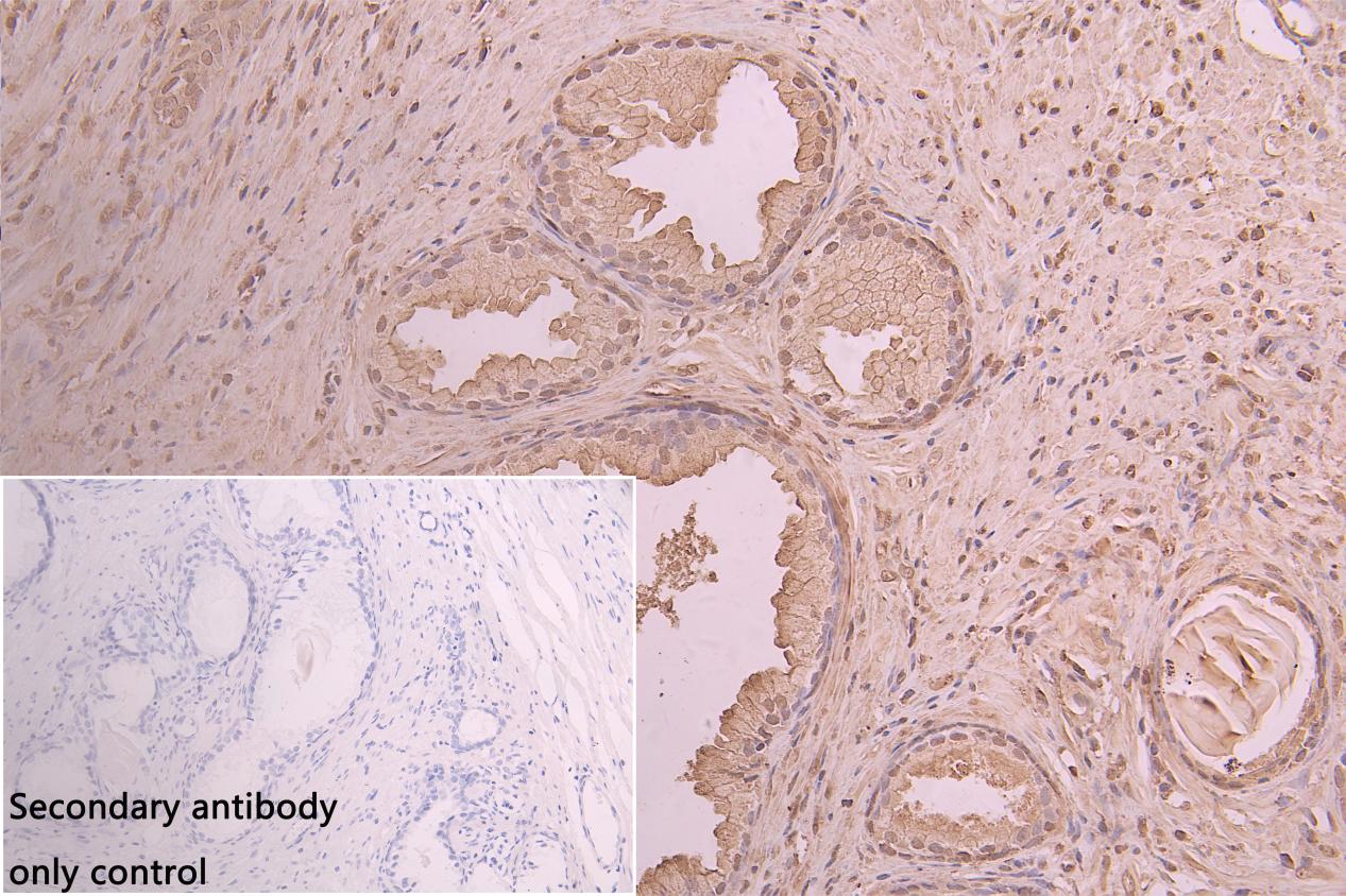
IHC image of CSB-PA001838ESR2HU diluted at 1:50 and staining in paraffin-embedded human prostate tissue performed on a Leica BondTM system. After dewaxing and hydration, antigen retrieval was mediated by high pressure in a citrate buffer (pH 6.0). Section was blocked with 10% normal goat serum 30min at RT. Then primary antibody (1% BSA) was incubated at 4C overnight. The primary is detected by a Goat anti-rabbit polymer IgG labeled by HRP and visualized using 0.05% DAB.Secondary antibody only control: uses 1% BSA instead of primary antibody |
|
|
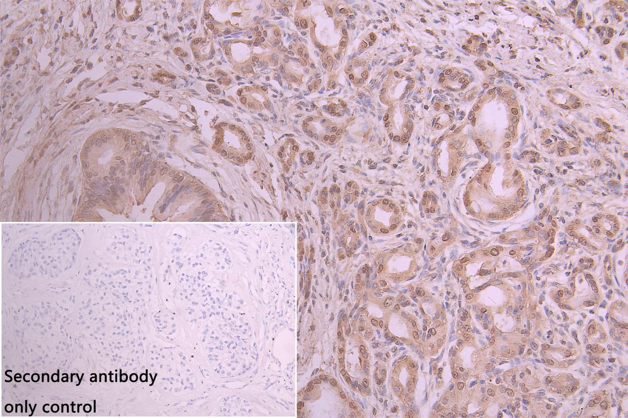
IHC image of CSB-PA001838ESR2HU diluted at 1:50 and staining in paraffin-embedded human pancreatic cancer performed on a Leica BondTM system. After dewaxing and hydration, antigen retrieval was mediated by high pressure in a citrate buffer (pH 6.0). Section was blocked with 10% normal goat serum 30min at RT. Then primary antibody (1% BSA) was incubated at 4C overnight. The primary is detected by a Goat anti-rabbit polymer IgG labeled by HRP and visualized using 0.05% DAB.Secondary antibody only control: uses 1% BSA instead of primary antibody |
|
|
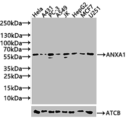
Western Blot Positive WB detected in: Hela whole cell lysate(20µg), A431 whole cell lysate(20µg), PC-3 whole cell lysate(20µg), A549 whole cell lysate(20µg), JK whole cell lysate(20µg), HepG2 whole cell lysate(20µg),MCF7 whole cell lysate(20µg),U251 whole cell lysate(20µg), All lanes: ANXA11 antibody at 1:1000 Secondary Goat polyclonal to rabbit IgG at 1/40000 dilution Predicted band size: 54,51,56,60 kDa Observed band size: 54,56 kDa Exposure time: 20s |
Product Guarantee and Expert Support
