
GSTM3 Antibody, Unconjugated, Rabbit, Polyclonal
Catalog Number:
CSB-PA009982LA01HU
- Images (9)


| Article Name: | GSTM3 Antibody, Unconjugated, Rabbit, Polyclonal |
| Biozol Catalog Number: | CSB-PA009982LA01HU |
| Supplier Catalog Number: | CSB-PA009982LA01HU |
| Alternative Catalog Number: | CSB-PA009982LA01HU-100UL |
| Manufacturer: | Cusabio |
| Host: | Rabbit |
| Category: | Antikörper |
| Application: | ELISA, IHC, WB |
| Species Reactivity: | Human, Mouse, Rat |
| Conjugation: | Unconjugated |
| Alternative Names: | brain GST antibody, brain type mu glutathione S transferase antibody, glutathione S alkyltransferase M3 antibody, glutathione S aryltransferase M3 antibody, glutathione S transferase M3 (brain) antibody, glutathione S transferase M3 antibody, Glutathione S transferase Mu 3 antibody, glutathione S transferase, Mu 3 antibody, Glutathione S-transferase Mu 3 antibody, GST class mu 3 antibody, GST class-mu 3 antibody, GST5 antibody, GSTB antibody, Gstm3 antibody, GSTM3-3 antibody, GSTM3_HUMAN antibody, GTM3 antibody, hGSTM3-3 antibody, MGC3310 antibody, MGC3704 antibody, S (hydroxyalkyl)glutathione lyase M3 antibody |
| Clonality: | Polyclonal |
| UniProt: | P21266 |
| Buffer: | Preservative: 0.02% sodium azide<br />Constituents: 50% Glycerol, 0.01M PBS, pH 7.4 |
| Purity: | Antigen Affinity Purified |
| Form: | Liquid |
| Target: | GSTM3 |
| Application Dilute: | Recommended dilution: WB:1:1000-1:5000, IHC:1:100-1:300 |
|
|
|
|
|
I |
|
|
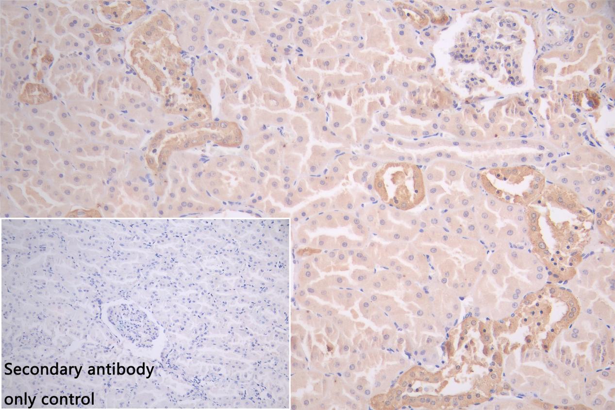
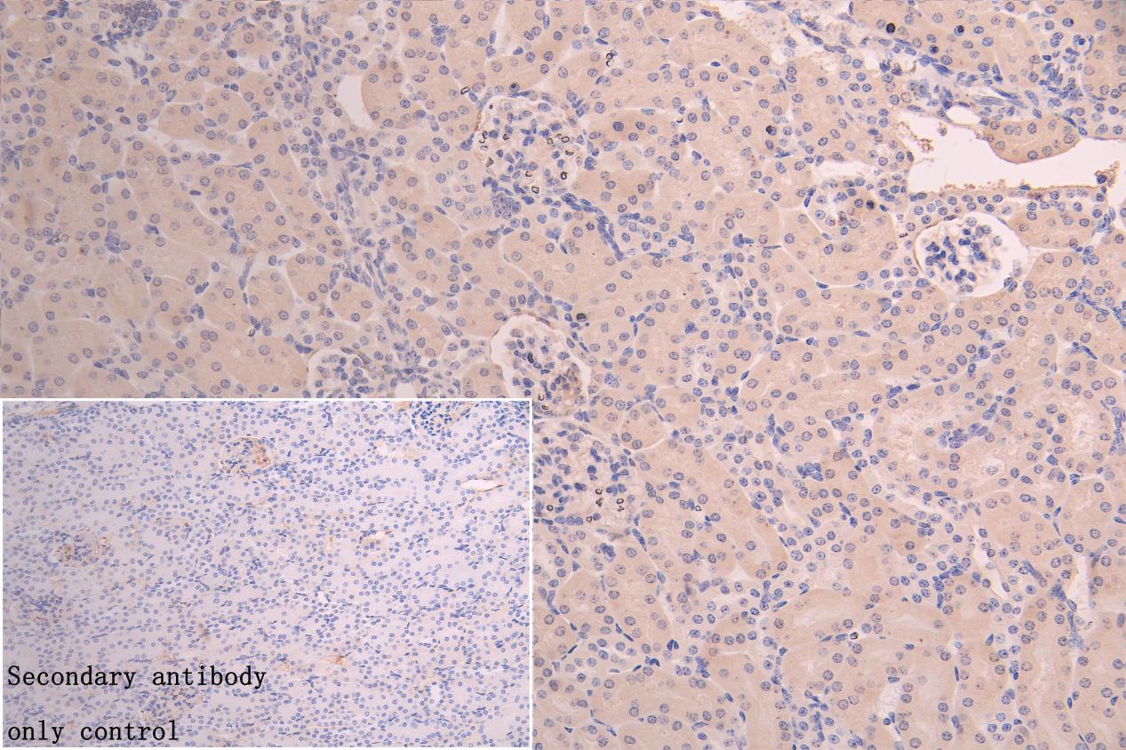
IHC image of CSB-PA009982LA01HU diluted at 1:100 and staining in paraffin-embedded human kidney tissue performed on a Leica BondTM system. After dewaxing and hydration, antigen retrieval was mediated by high pressure in a citrate buffer (pH 6.0). Section was blocked with 10% normal goat serum 30min at RT. Then primary antibody (1% BSA) was incubated at 4C overnight. The primary is detected by a Goat anti-rabbit polymer IgG labeled by HRP and visualized using 0.05% DAB. Secondary antibody only control: uses 1% BSA instead of primary antibody |
|
|
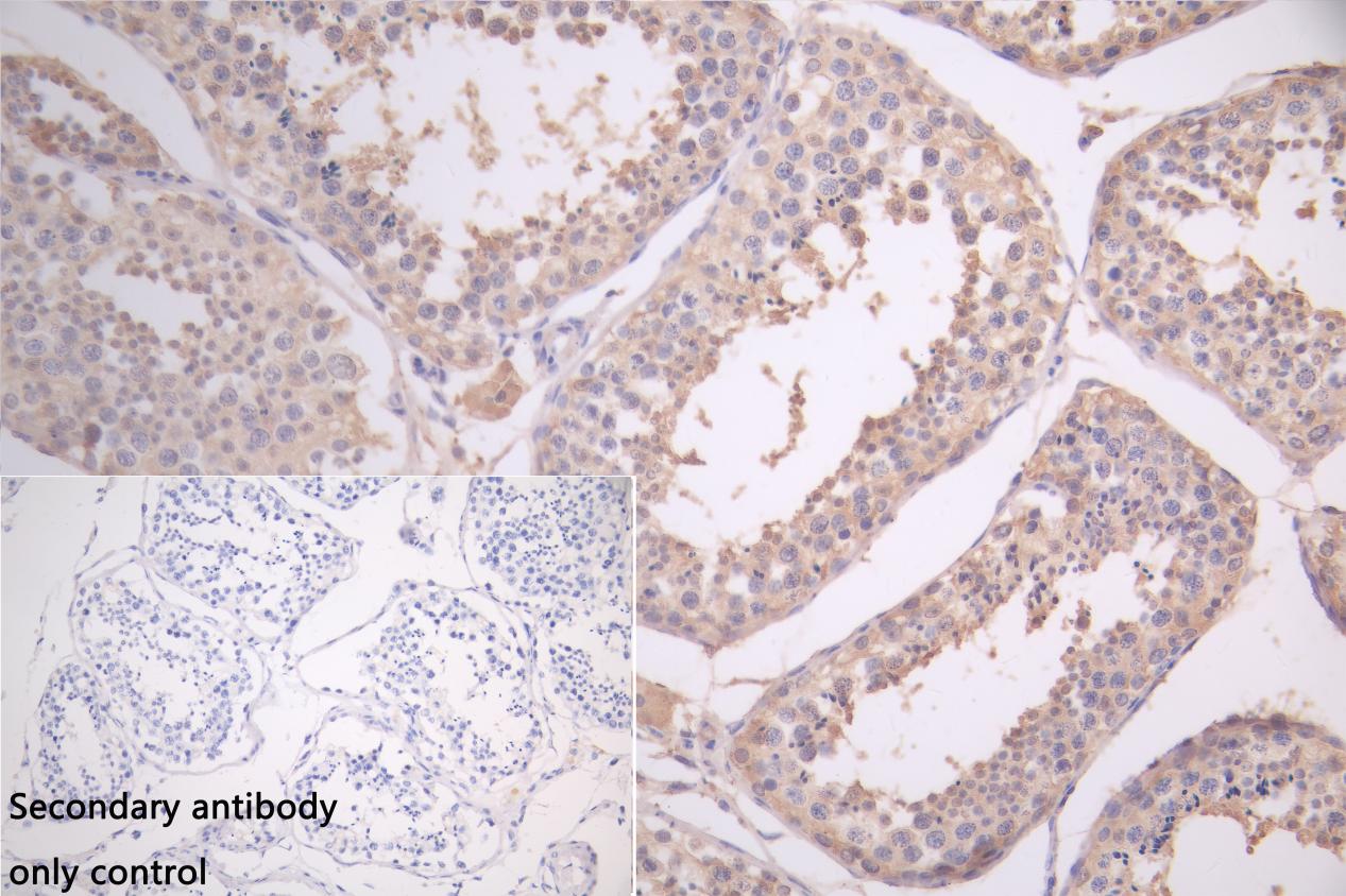
IHC image of CSB-PA009982LA01HU diluted at 1:100 and staining in paraffin-embedded human testis tissue performed on a Leica BondTM system. After dewaxing and hydration, antigen retrieval was mediated by high pressure in a citrate buffer (pH 6.0). Section was blocked with 10% normal goat serum 30min at RT. Then primary antibody (1% BSA) was incubated at 4C overnight. The primary is detected by a Goat anti-rabbit polymer IgG labeled by HRP and visualized using 0.05% DAB. Secondary antibody only control: uses 1% BSA instead of primary antibody |
|
|
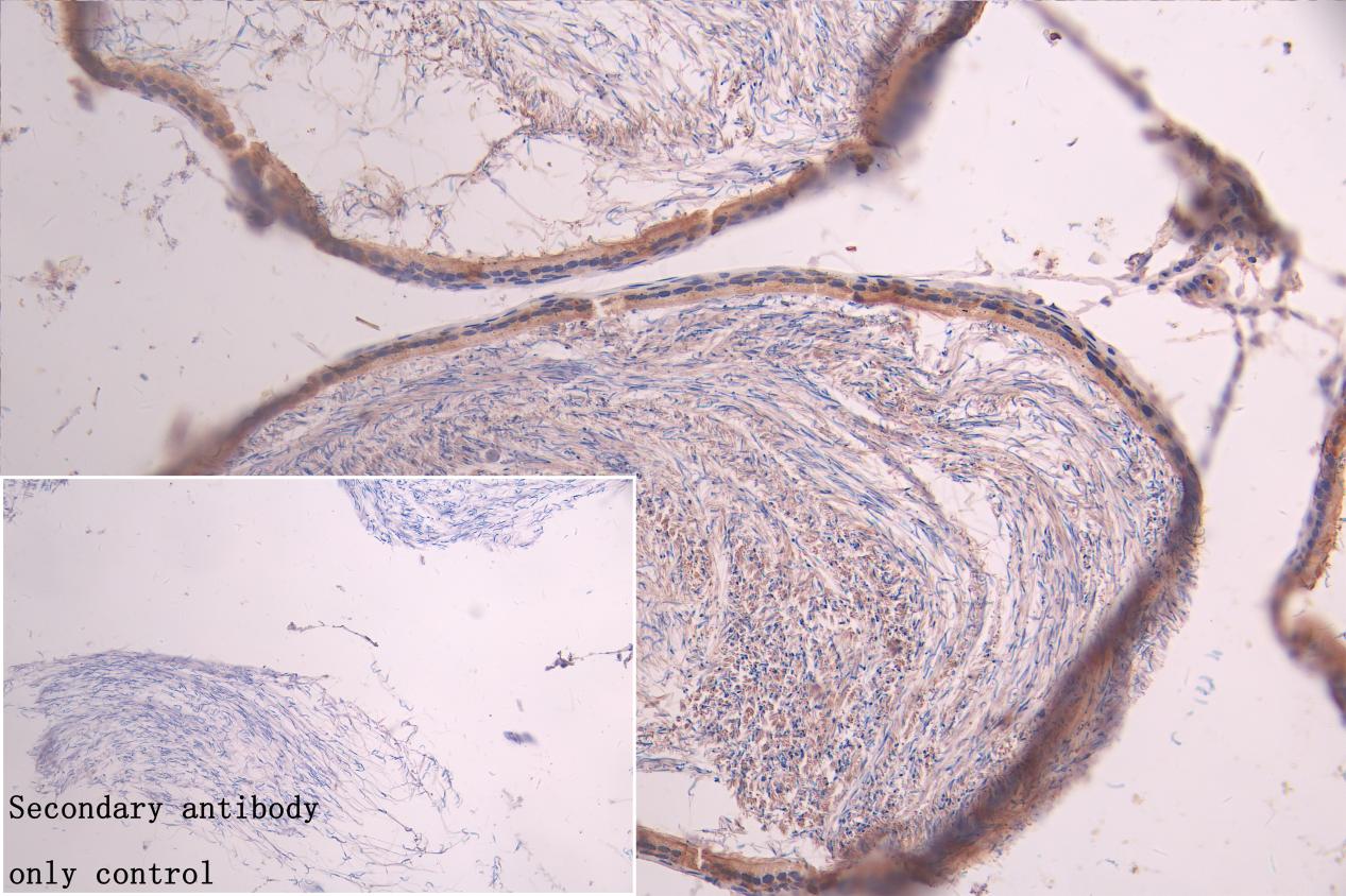
IHC image of CSB-PA009982LA01HU diluted at 1:100 and staining in paraffin-embedded rat testis tissue performed on a Leica BondTM system. After dewaxing and hydration, antigen retrieval was mediated by high pressure in a citrate buffer (pH 6.0). Section was blocked with 10% normal goat serum 30min at RT. Then primary antibody (1% BSA) was incubated at 4C overnight. The primary is detected by a Goat anti-rabbit polymer IgG labeled by HRP and visualized using 0.05% DAB.Secondary antibody only control: uses 1% BSA instead of primary antibody |
|
|
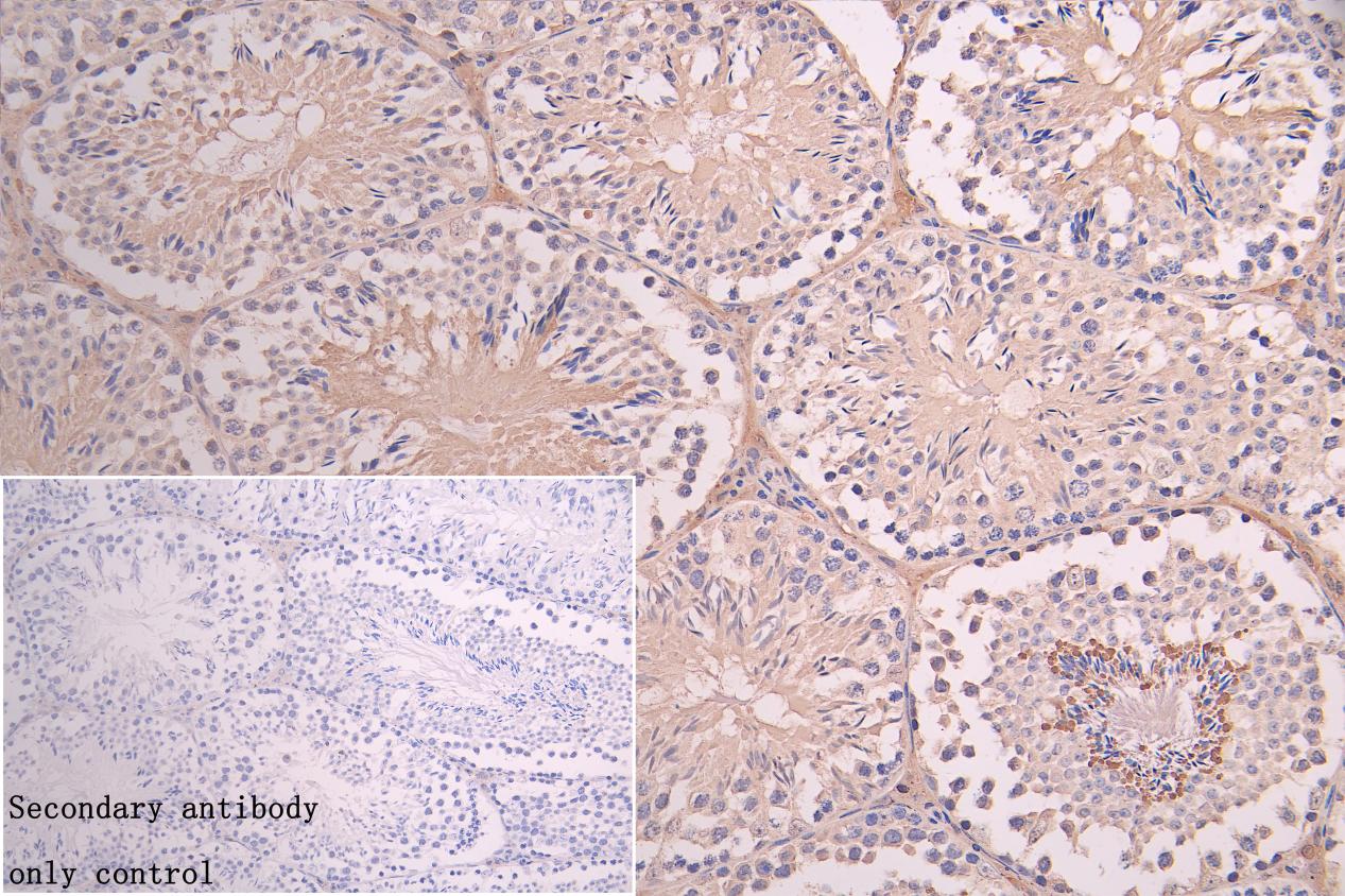
IHC image of CSB-PA009982LA01HU diluted at 1:100 and staining in paraffin-embedded mouse testis tissue performed on a Leica BondTM system. After dewaxing and hydration, antigen retrieval was mediated by high pressure in a citrate buffer (pH 6.0). Section was blocked with 10% normal goat serum 30min at RT. Then primary antibody (1% BSA) was incubated at 4C overnight. The primary is detected by a Goat anti-rabbit polymer IgG labeled by HRP and visualized using 0.05% DAB.Secondary antibody only control: uses 1% BSA instead of primary antibody |
|
|
IHC image of CSB-PA009982LA01HU diluted at 1:100 and staining in paraffin-embedded mouse kidney tissue performed on a Leica BondTM system. After dewaxing and hydration, antigen retrieval was mediated by high pressure in a citrate buffer (pH 6.0). Section was blocked with 10% normal goat serum 30min at RT. Then primary antibody (1% BSA) was incubated at 4C overnight. The primary is detected by a Goat anti-rabbit polymer IgG labeled by HRP and visualized using 0.05% DAB.Secondary antibody only control: uses 1% BSA instead of primary antibody |
|
|
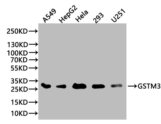
Western Blot Positive WB detected in: A549 whole cell lysate(20 µg), HepG2 whole cell lysate(20 µg), Hela whole cell lysate(20 µg), 293 whole cell lysate (20 µg), U251 whole cell lysate (20 µg) All lanes: GSTM3 antibody at 1:1000 Secondary Goat polyclonal to rabbit IgG at 1/50000 dilution Predicted band size: 27 kDa Observed band size: 27 kDa Exposure time:10s |
|
|
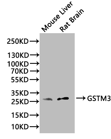
Western Blot Positive WB detected in: Mouse Liver tissue lysate(20µg),Rat Brain tissue lysate(20µg) All lanes: GSTM3 antibody at 1:1000 Secondary Goat polyclonal to rabbit IgG at 1/50000 dilution Predicted band size: 27 kDa Observed band size: 27 kDa Exposure time:120s |
Product Guarantee and Expert Support
