
LIPA Antibody, Unconjugated, Rabbit, Polyclonal
Catalog Number:
CSB-PA012972EA01HU
- Images (9)


| Article Name: | LIPA Antibody, Unconjugated, Rabbit, Polyclonal |
| Biozol Catalog Number: | CSB-PA012972EA01HU |
| Supplier Catalog Number: | CSB-PA012972EA01HU |
| Alternative Catalog Number: | CSB-PA012972EA01HU-100UL |
| Manufacturer: | Cusabio |
| Host: | Rabbit |
| Category: | Antikörper |
| Application: | ELISA, IF, IHC, WB |
| Species Reactivity: | Human |
| Conjugation: | Unconjugated |
| Alternative Names: | Acid cholesteryl ester hydrolase antibody, CESD antibody, cholesterol ester hydrolase antibody, cholesterol ester storage disease antibody, Cholesteryl esterase antibody, Hydrolase deficiency antibody, LAL antibody, LAL deficiency cholesterol ester antibody, LICH_HUMAN antibody, lipA antibody, LIPA deficiency antibody, Lipase A antibody, lipase A, lysosomal acid, cholesterol esterase antibody, lysosomal acid lipase antibody, lysosomal acid lipase deficiency antibody, Lysosomal acid lipase/cholesteryl ester hydrolase antibody, Sterol esterase antibody |
| Clonality: | Polyclonal |
| UniProt: | P38571 |
| Buffer: | Preservative: 0.02% sodium azide Constituents: PBS containing 50% glycerol pH 7.3 |
| Purity: | Antigen Affinity Purified |
| Form: | Liquid |
| Target: | LIPA |
| Application Dilute: | Recommended dilution: WB:1:500-1:2000,IHC:1:20-1:200,IF:1:100-1:100 |
|
|
Immunohistochemistry of paraffi |
|
|
|
|
|
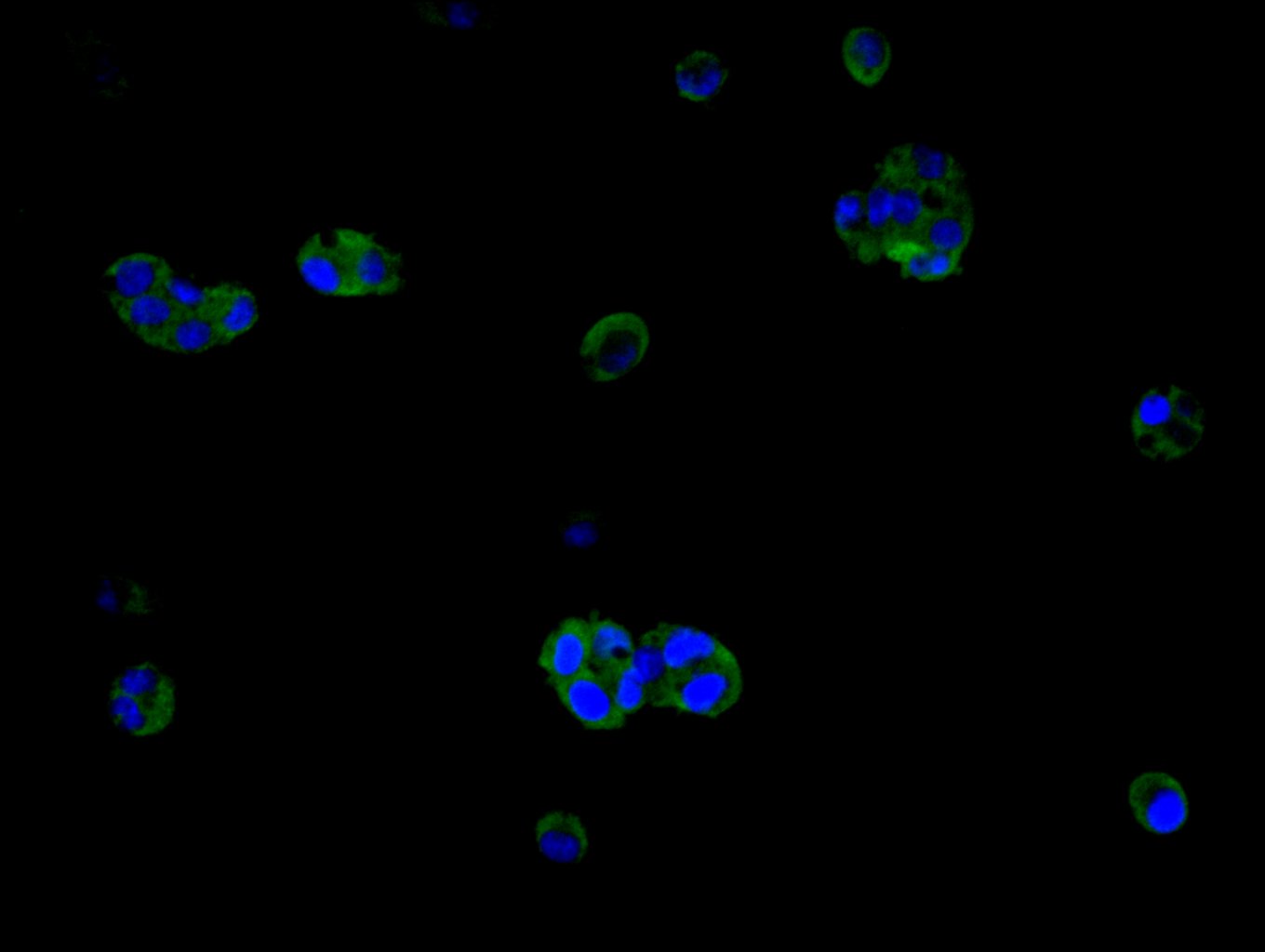
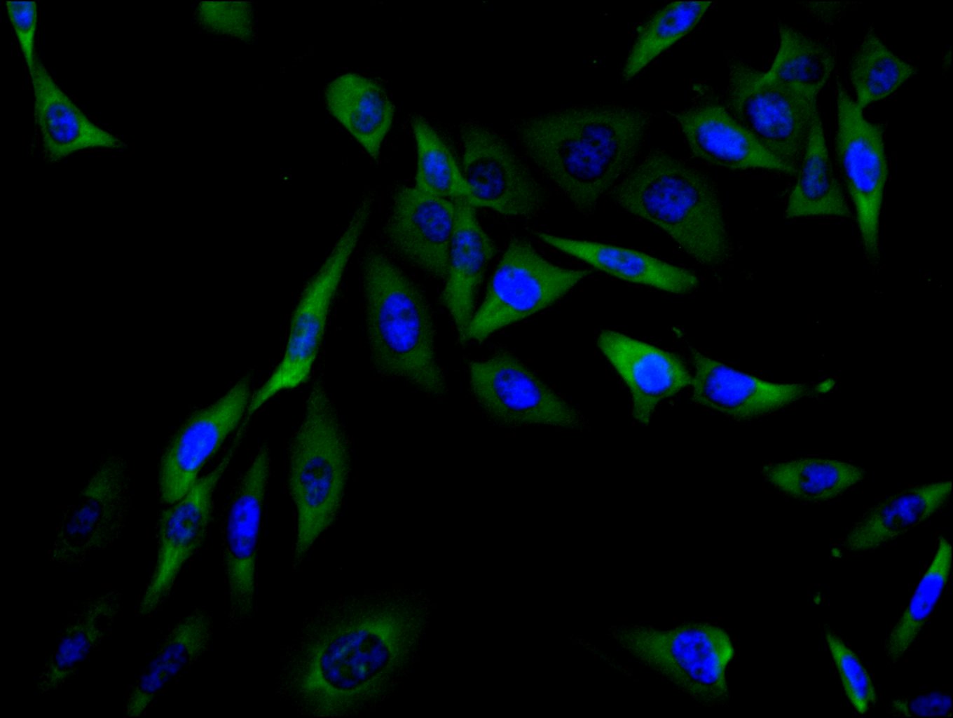
Immunofluorescence staining of HepG2 cell with CSB-PA012972EA01HU at 1:15, counter-stained with DAPI. The cells were fixed in 4% formaldehyde and and permeated by 0.2% TritonX-100 for 15 min. Then 10% normal goat serum to block non-specific protein-protein interactions . The cells were then incubated with the antibody overnight at 4°C. The secondary antibody was Alexa Fluor 488-congugated AffiniPure Goat Anti-Rabbit IgG(H+L). |
|
|
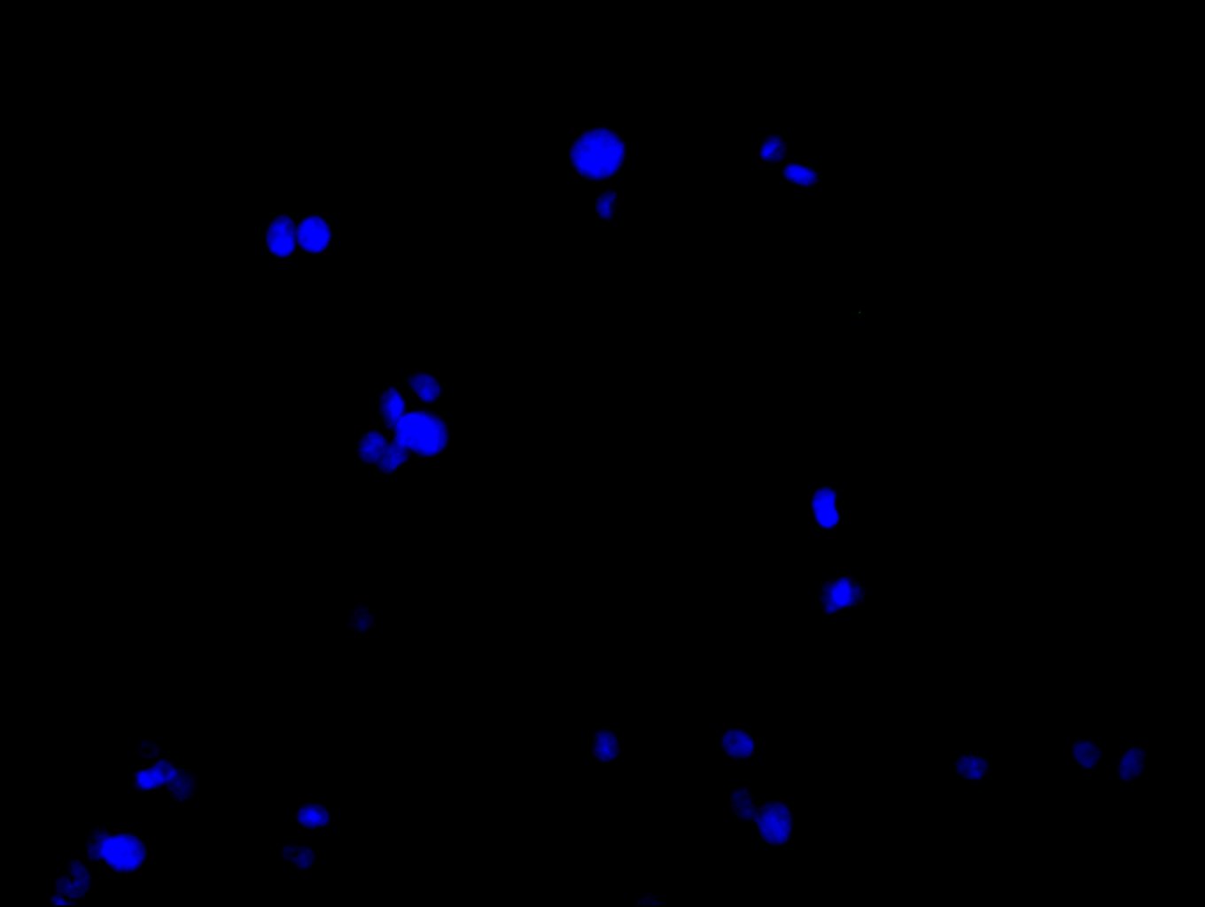
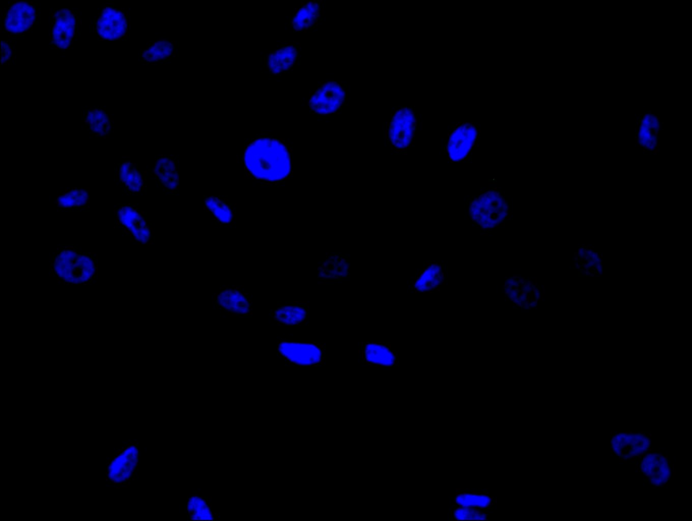
Immunofluorescence staining of HepG2 cell with 5% goat serum, counter-stained with DAPI. The cells were fixed in 4% formaldehyde and blocked in 10% normal Goat Serum. The cells were then incubated with the antibody overnight at 4C. The secondary antibody was Alexa Fluor 488-congugated AffiniPure Goat Anti-Rabbit IgG(H+L). |
|
|
Immunofluorescence staining of PC-3 cell with CSB-PA012972EA01HU at 1:15, counter-stained with DAPI. The cells were fixed in 4% formaldehyde and and permeated by 0.2% TritonX-100 for 15 min. Then 10% normal goat serum to block non-specific protein-protein interactions . The cells were then incubated with the antibody overnight at 4°C. The secondary antibody was Alexa Fluor 488-congugated AffiniPure Goat Anti-Rabbit IgG(H+L). |
|
|
Immunofluorescence staining of PC-3 cell with 5% goat serum, counter-stained with DAPI. The cells were fixed in 4% formaldehyde and blocked in 10% normal Goat Serum. The cells were then incubated with the antibody overnight at 4C. The secondary antibody was Alexa Fluor 488-congugated AffiniPure Goat Anti-Rabbit IgG(H+L). |
|
|
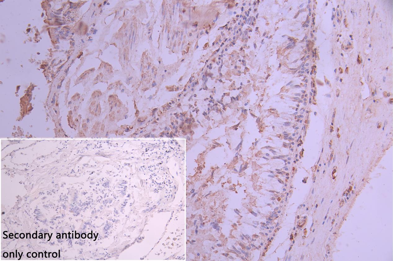
IHC image of CSB-PA012972EA01HU diluted at 1:30 and staining in paraffin-embedded human Lung tissue performed on a Leica BondTM system. After dewaxing and hydration, antigen retrieval was mediated by high pressure in a citrate buffer (pH 6.0). Section was blocked with 10% normal goat serum 30min at RT. Then primary antibody (1% BSA) was incubated at 4C overnight. The primary is detected by a Goat anti-rabbit polymer IgG labeled by HRP and visualized using 0.05% DAB. Secondary antibody only control: uses 1% BSA instead of primary antibody |
|
|
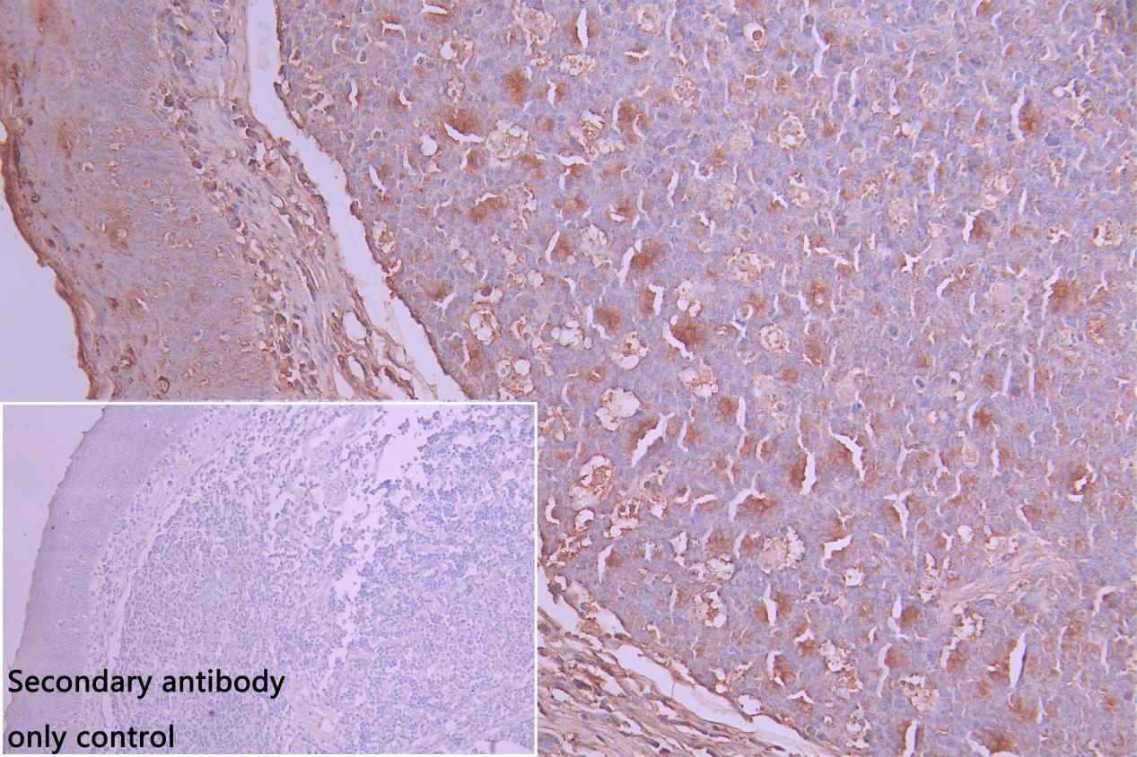
IHC image of CSB-PA012972EA01HU diluted at 1:30 and staining in paraffin-embedded human tonsil tissue performed on a Leica BondTM system. After dewaxing and hydration, antigen retrieval was mediated by high pressure in a citrate buffer (pH 6.0). Section was blocked with 10% normal goat serum 30min at RT. Then primary antibody (1% BSA) was incubated at 4C overnight. The primary is detected by a Goat anti-rabbit polymer IgG labeled by HRP and visualized using 0.05% DAB. Secondary antibody only control: uses 1% BSA instead of primary antibody |
|
|
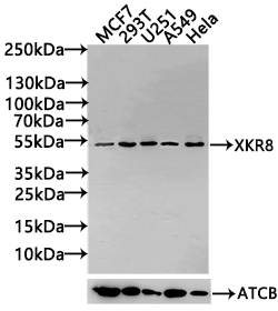
Western Blot Positive WB detected in: MCF7 whole cell lysate(30µg), 293T whole cell lysate(30µg), U251 whole cell lysate(30µg), A549 whole cell lysate(30µg), Hela whole cell lysate(30µg) All lanes: LIPA antibody at 1:1000 Secondary Goat polyclonal to rabbit IgG at 1/20000 dilution Predicted band size:46,40 kDa Observed band size: 46 kDa Exposure time: 10s |
Product Guarantee and Expert Support
