
NQO2 Antibody, Unconjugated, Rabbit, Polyclonal
Catalog Number:
CSB-PA016040LA01HU
- Images (9)


| Article Name: | NQO2 Antibody, Unconjugated, Rabbit, Polyclonal |
| Biozol Catalog Number: | CSB-PA016040LA01HU |
| Supplier Catalog Number: | CSB-PA016040LA01HU |
| Alternative Catalog Number: | CSB-PA016040LA01HU-100UL |
| Manufacturer: | Cusabio |
| Host: | Rabbit |
| Category: | Antikörper |
| Application: | ELISA, IF, IHC, WB |
| Species Reactivity: | Human |
| Conjugation: | Unconjugated |
| Alternative Names: | DHQV antibody, DIA6 antibody, EC 1.10.99.2 antibody, MGC94180 antibody, NAD(P)H dehydrogenase quinone 2 antibody, NAD(P)H menadione oxidoreductase 1 dioxin inducible 2 antibody, NAD(P)H menadione oxidoreductase 2 dioxin inducible antibody, NMOR2 antibody, NQO 2 antibody, NQO2 antibody, NQO2_HUMAN antibody, NRH dehydrogenase [quinone] 2 antibody, NRH dehydrogenase antibody, NRH:quinone oxidoreductase 2 antibody, OTTHUMP00000015948 antibody, OTTHUMP00000015949 antibody, OTTHUMP00000015953 antibody, Ox 2 antibody, Ox2 antibody, QR2 antibody, Quinone antibody, Quinone reductase 2 antibody, Ribosyldihydronicotinamide dehydrogenase [quinone] antibody, Ribosyldihydronicotinamide dehydrogenase antibody |
| Clonality: | Polyclonal |
| UniProt: | P16083 |
| Buffer: | Preservative: 0.02% sodium azide<br />Constituents: PBS containing 50% glycerol pH 7.3 |
| Purity: | Antigen Affinity Purified |
| Form: | Liquid |
| Target: | NQO2 |
| Application Dilute: | Recommended dilution: WB:1:1000-1:4000, IHC:1:100-1:300, IF:1:20-1:100 |
|
|
|
|
|
Immunohistochemistry of paraffin-embedded human brain tissue using CSB-PA016040LA01HU at dilution of 1:100 |
|
|
Western Blot Positive WB detected in: Hela whole cell lysate, K562 whole cell lysate, HepG2 whole cell lysate, JK whole cell lysate, A549 whole cell lysate, MCF7 whole cell lysate, ntera2 whole cell lysate All lanes:NQO2 Antibody at 1:1000 Secondary Goat polyclonal to rabbit IgG at 1/50000 dilution Predicted band size:26 kDa Observed band size:26 kDa |
|
|
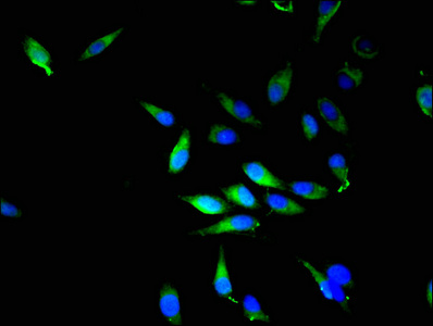
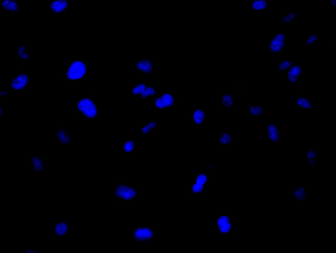
Immunofluorescence stainingof Hela cell with CSB-PA016040LA01HU at 1:20, counter-stained with DAPI. The cells were fixed in 4% formaldehyde and blocked in 10% normal Goat Serum. The cells were then incubated with the antibody overnight at 4C. The secondary antibody was Alexa Fluor 488-congugated AffiniPure Goat Anti-Rabbit IgG(H+L). |
|
|
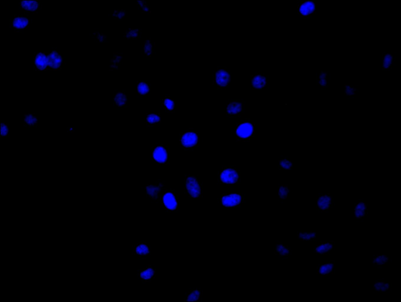
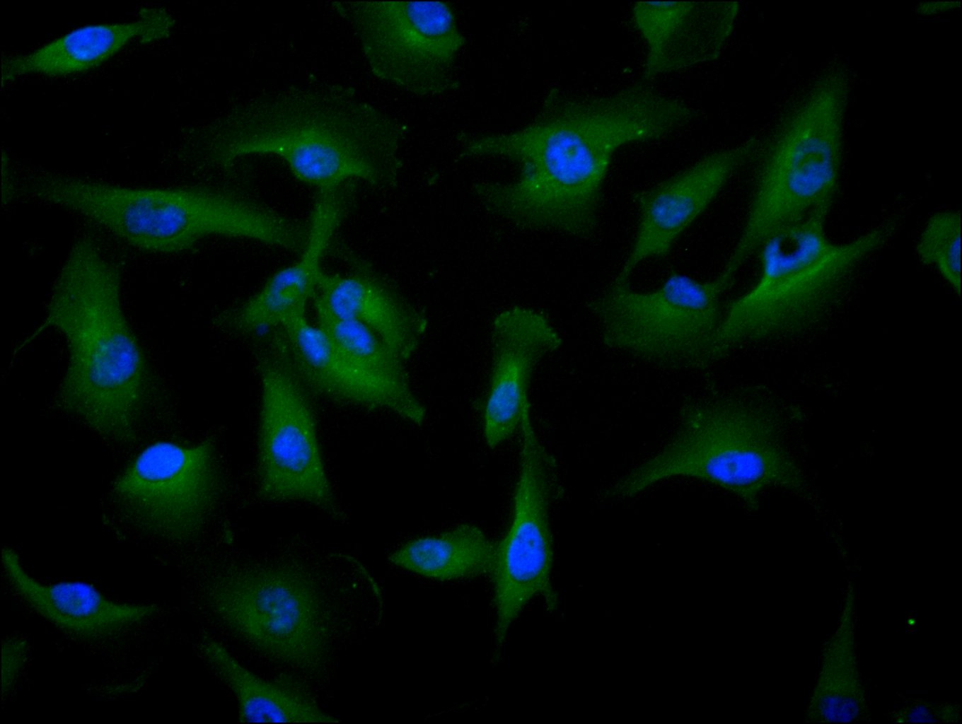
Immunofluorescence staining of Hela cell with 5% goat serum, counter-stained with DAPI. The cells were fixed in 4% formaldehyde and blocked in 10% normal Goat Serum. The cells were then incubated with the antibody overnight at 4C. The secondary antibody was Alexa Fluor 488-congugated AffiniPure Goat Anti-Rabbit IgG(H+L). |
|
|
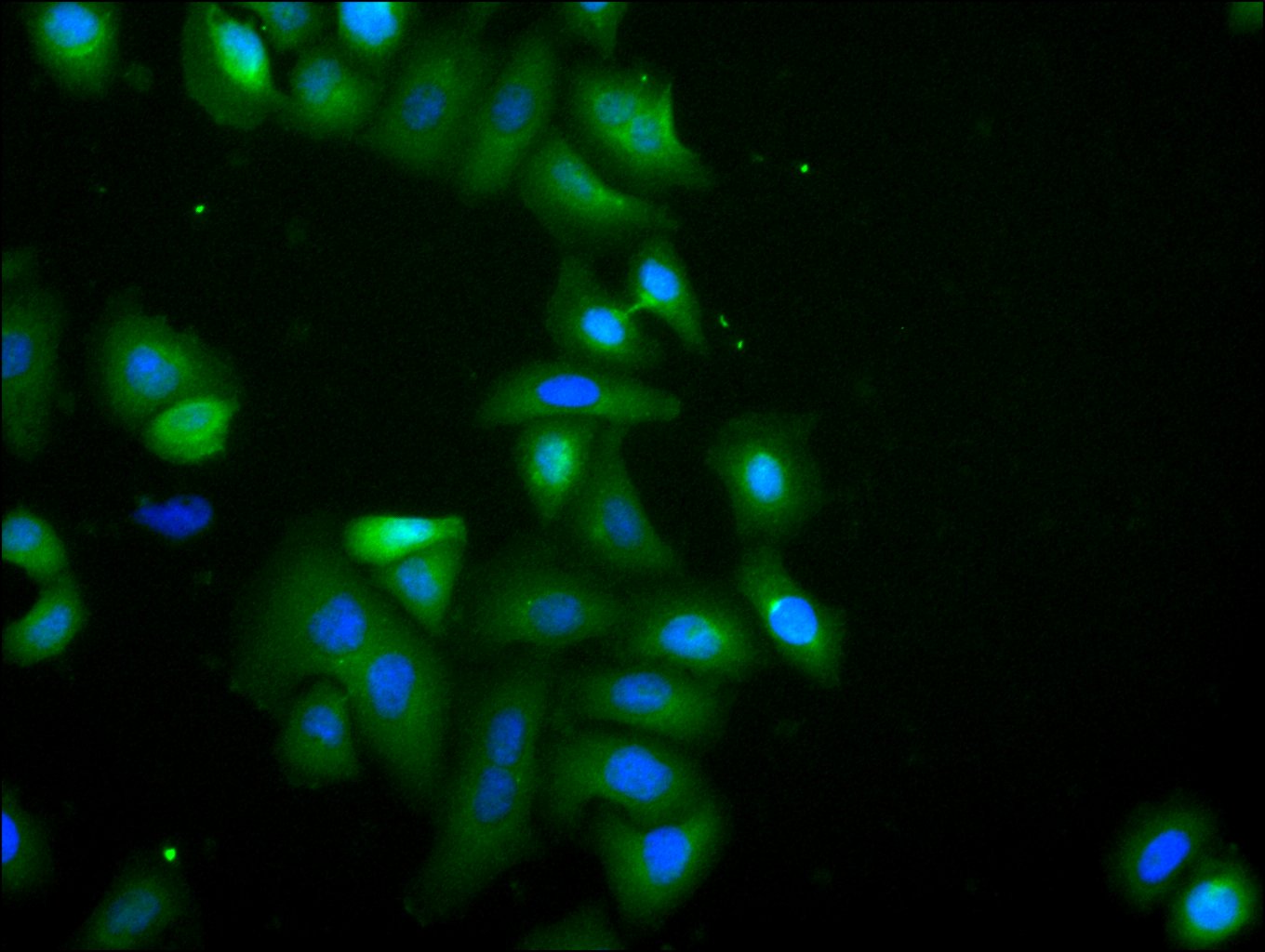
Immunofluorescence stainingof U251 cell with CSB-PA016040LA01HU at 1:20, counter-stained with DAPI. The cells were fixed in 4% formaldehyde and blocked in 10% normal Goat Serum. The cells were then incubated with the antibody overnight at 4C. The secondary antibody was Alexa Fluor 488-congugated AffiniPure Goat Anti-Rabbit IgG(H+L). |
|
|
Immunofluorescence staining of Hela cell with 5% goat serum, counter-stained with DAPI. The cells were fixed in 4% formaldehyde and blocked in 10% normal Goat Serum. The cells were then incubated with the antibody overnight at 4C. The secondary antibody was Alexa Fluor 488-congugated AffiniPure Goat Anti-Rabbit IgG(H+L). |
|
|
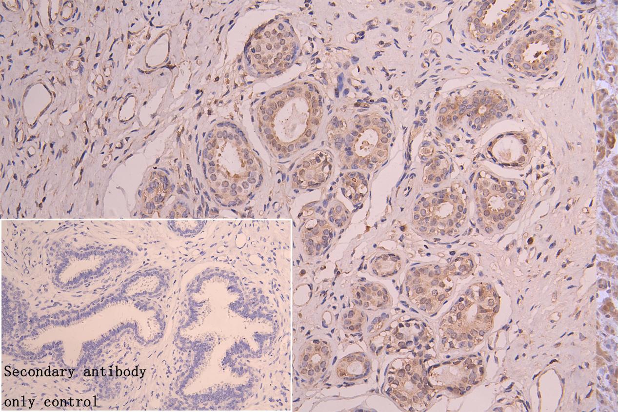
IHC image of CSB-PA016040LA01HU diluted at 1:150 and staining in paraffin-embedded human breast tissue performed on a Leica BondTM system. After dewaxing and hydration, antigen retrieval was mediated by high pressure in a citrate buffer (pH 6.0). Section was blocked with 10% normal goat serum 30min at RT. Then primary antibody (1% BSA) was incubated at 4C overnight. The primary is detected by a Goat anti-rabbit polymer IgG labeled by HRP and visualized using 0.05% DAB. Secondary antibody only control: uses 1% BSA instead of primary antibody |
|
|
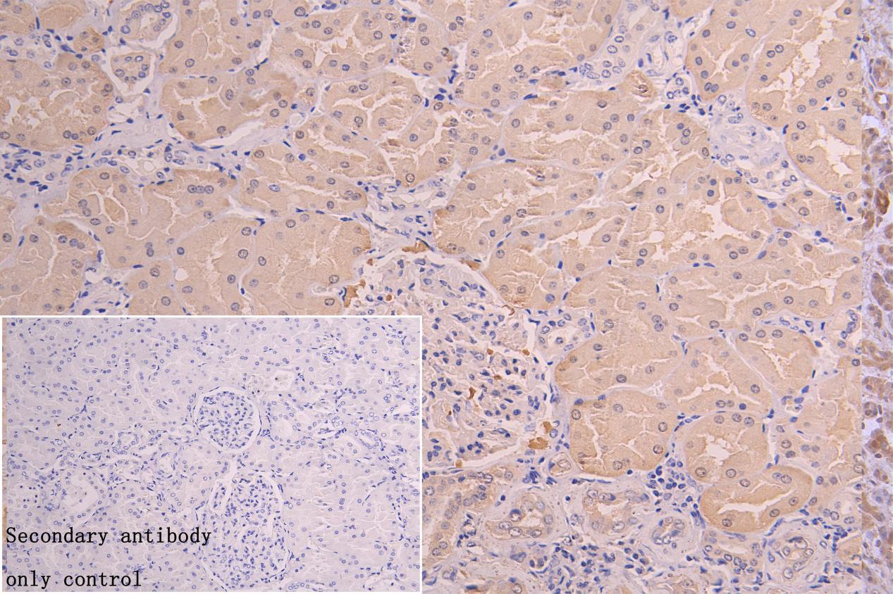
IHC image of CSB-PA016040LA01HU diluted at 1:150 and staining in paraffin-embedded human kidney tissue performed on a Leica BondTM system. After dewaxing and hydration, antigen retrieval was mediated by high pressure in a citrate buffer (pH 6.0). Section was blocked with 10% normal goat serum 30min at RT. Then primary antibody (1% BSA) was incubated at 4C overnight. The primary is detected by a Goat anti-rabbit polymer IgG labeled by HRP and visualized using 0.05% DAB. Secondary antibody only control: uses 1% BSA instead of primary antibody |
Product Guarantee and Expert Support
