DYKDDDDK Tag (Anti-FLAG) Antibody, Rabbit, Polyclonal
Biozol Catalog Number:
ROC-600-401-383
Supplier Catalog Number:
600-401-383
Alternative Catalog Number:
ROC-600-401-383
Manufacturer:
Rockland Immunochemicals
Host:
Rabbit
Category:
Antikörper
Application:
ELISA, WB
Immunogen:
This antibody was purified from whole rabbit serum prepared by repeated immunizations with the Enterokinase Cleavage Site (ECS) peptide DYKDDDDK (Asp-Tyr-Lys-Asp-Asp-Asp-Asp-Lys) conjugated to KLH using maleimide. This antibody reacts with FLAG(TM) conjugated proteins.
Conjugation:
Unconjugated
Alternative Names:
rabbit antibody for the detection of FLAG(TM) conjugated proteins, rabbit anti DYKDDDDK
Clonality:
Polyclonal
Concentration:
1.09 by UV absorbance at 280 nm
Buffer:
0.02 M Potassium Phosphate, 0.15 M Sodium Chloride, pH 7.2
This antibody is optimally suited for monitoring the expression of FLAG(TM) tagged fusion proteins. As such, this antibody can be used to identify fusion proteins containing the FLAG(TM) epitope. The antibody recognizes the epitope tag fused to either the amin
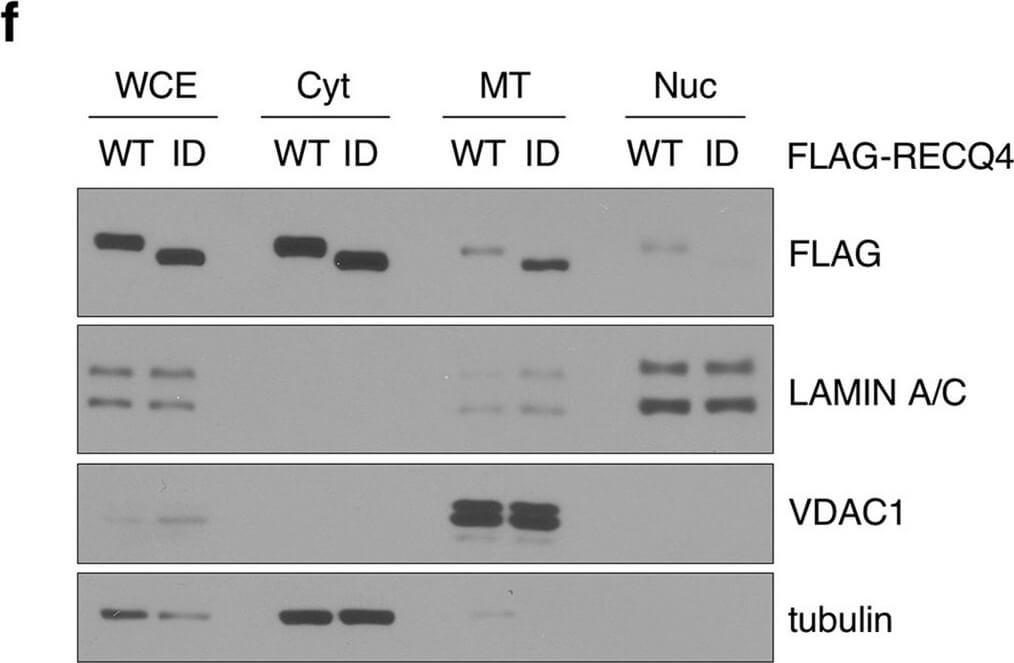
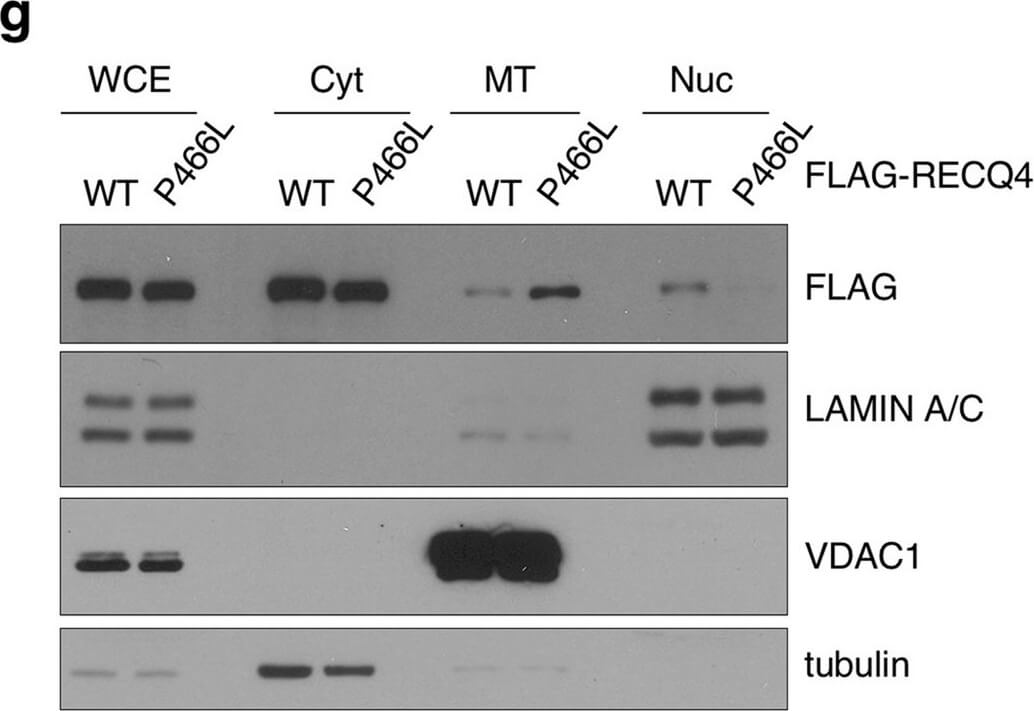
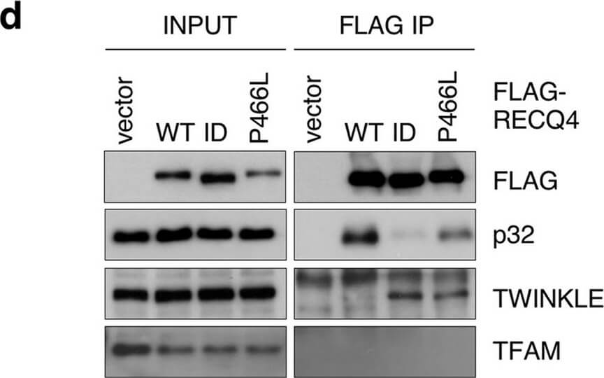
The P466L clinical mutation leads to RECQ4 mitochondrial accumulation. (a) Schematic of human RECQ4 WT, ID and P466L mutant proteins, including the SLD2 (green) and conserved SF2 helicase domains (yellow). (b) Western blot analysis of RECQ4 in WCEs and chromatin-bound (CB) fractions prepared from HEK293 WT or RECQ4 knockdown (KD) HEK293 cells generated by CRISPR technology. Tubulin is used as a loading control. (c) The effects of stable RECQ4 KD shown in (b) and complementation using FLAG-RECQ4 on cell growth as measured by crystal violet cell proliferation assays. Each value represents meanstandard deviation of 3 independent biological experiments, each with 3 triplicate reactions. (d) Western blot analysis for the presence of WT and mutant FLAG-RECQ4, p32, TWINKLE and TFAM in WCE (left) and immunoprecipitated (IP) with FLAG-RECQ4 complexes (right) in WCEs prepared from stable RECQ4 KD HEK293 cells expressing FLAG-RECQ4 WT, ID or P466L mutant. (e) gDNA levels relative to mtDNA in WCE (left) and MT (right) prepared from stable RECQ4 KD HEK293 cells expressing FLAG-RECQ4 WT, ID or P466L mutant. (f) Western blot analysis of stable RECQ4 KD HEK293 cells expressing FLAG-RECQ4 WT or ID mutant in WCEs and Cyt, MT, and fractions. Tubulin, VDAC1, and lamin A/C are loading and fractionation controls for Cyt, MT, and Nuc fractions, respectively. (g) Western blot analysis of RECQ4 in WCEs and Cyt, MT, and Nuc fractions prepared from stable RECQ4 KD HEK293 cells expressing FLAG-RECQ4 WT or P466L mutant. (h) Representative images showing immunofluorescent staining of FLAG-RECQ4 (green) in stable RECQ4 KD HEK293 cells expressing indicated WT and mutant FLAG-RECQ4 proteins. Mitotracker (red) was used to detect mitochondria, and DAPI (blue) was used to detect nuclei. Figure provided by CiteAb. Source: Sci Rep, PMID: 33046774.
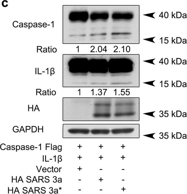
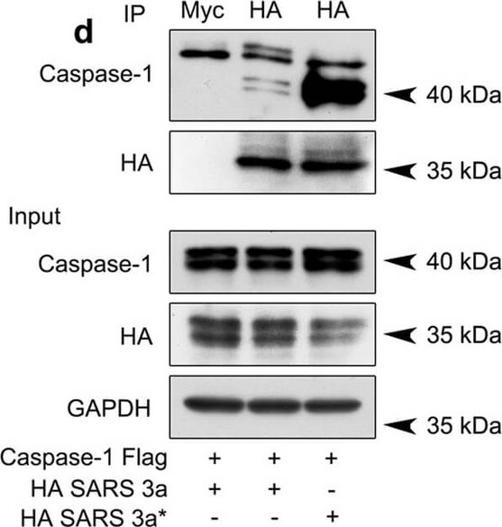
SARS 3a induces NLRP3 inflammasome activation by multiple mechanisms. A) Immunoblot analysis of the pro- and cleaved forms of caspase-1 and IL-1beta after reconstitution of inflammasome in HEK 293T cells transfected with SARS 3a with or without NEK7 shRNA. B) Immunoblot analysis of the pro- and cleaved forms of caspase-1 and IL-1beta after reconstitution of inflammasome and transfection with SARS 3a or SARS 3a C133A. C) Immunoblot analysis of the pro- and cleaved forms of caspase-1 and IL-1beta after co-transfection with caspase-1, IL-1beta, and SARS 3a or SARS 3a C133A. D) Immunoprecipitation analysis of interaction between SARS 3a or SARS 3a C133A and caspase-1. All western blot data are representative of two or three independent experiments Figure provided by CiteAb. Source: Cell Death Dis, PMID: 30185776.
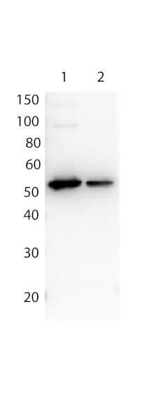
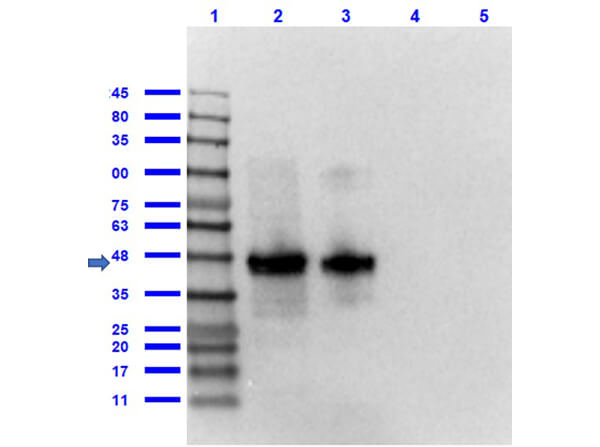
Affinity Purified Antibody to detect FLAG conjugated proteins detects both C terminal linked and N terminal linked FLAG tagged recombinant proteins by western blot. This antibody was used at a dilution of 1:1,000 to detect 0.1 µg of recombinant protein containing either the FLAG epitope tag linked at the carboxy (C), Lane 2, or the amino (N), Lane 1, terminus of the recombinant protein. A 4-20% gradient gel was used to resolve the protein by SDS-PAGE. The protein was transferred to nitrocellulose using standard methods. After blocking, the membrane was probed with the primary antibody overnight at 4C followed by washes and reaction with a 1:40,000 dilution of HRP conjugate
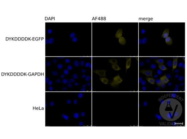
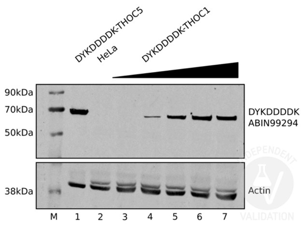
Immunofluorescence of the antibody for the detection of FLAG conjugated proteins. Cells: HeLa cells transiently expressing DYKDDDDK-tagged EGFP (top row), or GAPDH (middle row), or mock-transfected HeLa cells (bottom row). Fixation: 4% PFA for 20-30 min at RT. Primary Antibody: DYKDDDDK Tag antibody diluted 1:200 overnight at 4C. Secondary Antibody: chicken anti-rabbit AF488 antibody. Counterstain: DAPI for 10min at RT. Staining: DAPI (left column), DYKDDDDK Tag/AF488 (middle column), Merged DAPI and tag staining (right column). Independently Validated byantibodies-online GmbH (p/n ABIN1043869/ ABIN99294) courtesy ofUniversity of Bern.
* VAT and and shipping costs not included. Errors and price changes excepted