
Mcl-1 Antibody, Rabbit, Polyclonal
Catalog Number:
ROC-600-401-394
- Images (20)

| Article Name: | Mcl-1 Antibody, Rabbit, Polyclonal |
| Biozol Catalog Number: | ROC-600-401-394 |
| Supplier Catalog Number: | 600-401-394 |
| Alternative Catalog Number: | ROC-600-401-394 |
| Manufacturer: | Rockland Immunochemicals |
| Host: | Rabbit |
| Category: | Antikörper |
| Application: | ELISA, WB |
| Species Reactivity: | Mouse |
| Immunogen: | This affinity purified antibody was purified from whole rabbit serum prepared by repeated immunizations with a synthetic peptide corresponding to an internal region of mouse Mcl-1 conjugated to Keyhole Limpet Hemocyanin (KLH). |
| Conjugation: | Unconjugated |
| Alternative Names: | rabbit anti-Mcl-1 antibody, Mcl1, Mcl 1, Bcl 2 related protein EAT/mcl1 antibody, Bcl2 related antibody, EAT antibody, Induced myeloid leukemia cell differentiation protein Mcl-1 antibody |
| Application Dilute: | ELISA: 1:10,000 - 1:50,000, WB: 1:10,000 |
| Application Notes: | Anti-Mcl-1 Antibody has been tested by ELISA and western blot and is suitable for immunoprecipitation. This antibody detects mouse Mcl-1 and is not expected to cross react with the human sequence. Cross reactivity with Mcl-1 from other sources is unknown |
|
|
|
|
|
|
|
|
|
|
|
|
|
|
|
|
|
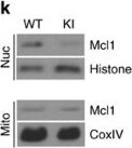
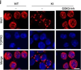
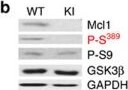
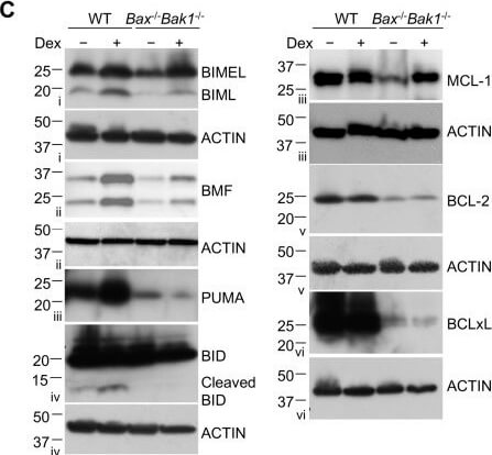
Inactivation of GSK3beta by p38 MAPK promotes accumulation of the prosurvival factor Mcl-1.(a,b) WT and GSK3beta-KI B cells were activated for two days and the expression of beta-catenin (a), Mcl-1, P-S389 GSK3beta and total GSK3beta (b) were examined by western blotting. GAPDH is shown as a loading control. (c) WT and GSK3beta-KI B cells were activated as in a and Bclx, Bcl2, Bid, Bax and Bad levels were examined by western blotting. (d) Western blot analysis for PUMA in irradiated WT thymocytes (5Gy) and activated WT and GSK3beta-KI B cells. (e,f) B cells activated as in a were stained with MitoTracker (e) or TMRE (f) and analysed by flow cytometry. The number represents the percentage of cells within the gate. (g) WT and GSK3beta-KI B cells were examined by western blotting for the expression of cleaved caspase-3 (activated caspase-3), full length RIPK1 (RIPK1) and cleaved RIPK1 (cleaved RIPK1). (h) WT and GSK3beta-KI B cells were activated, after 2 days Nectrostatin-1s (Nec-1s) was added and cell viability was determined by cell counting 24h later (n=3). (i) WT and GSK3beta-KI B cells were activated for 3 days and phospho-MLKL was examined by western blotting. (j) WT and GSK3beta-KI B cells in the presence or absence of the GSK3 inhibitor (GSK3-Inh) were activated and examined by immunostaining and confocal microscopy for Mcl-1 (red) and TOPRO nuclear stain (blue). Scale bar, 3µm. (k) Mcl-1 levels in nuclear (Nuc) and mitochondrial (Mito) extracts from activated WT and GSK3beta-KI B cells were determined by western blot analysis. Histone and CoxIV (Complex IV) were used as loading controls. (l) WT and GSK3beta-KI B cells were transduced with either an empty retrovirus (E), a retrovirus expressing wildtype Mcl-1 (Mcl) or a retrovirus expressing a Ser140Ala mutant of Mcl-1 (mMcl). Three days after activation cell viability was determined by cell counting. (n=3) and *P value<0.05 as determined by t- test (h) or one-way ANOVA (l). Data are representative of three or more independent experiments. Figure provided by CiteAb. Source: Nat Commun, PMID: 26822034. |
|
|
Inactivation of GSK3beta by p38 MAPK promotes accumulation of the prosurvival factor Mcl-1.(a,b) WT and GSK3beta-KI B cells were activated for two days and the expression of beta-catenin (a), Mcl-1, P-S389 GSK3beta and total GSK3beta (b) were examined by western blotting. GAPDH is shown as a loading control. (c) WT and GSK3beta-KI B cells were activated as in a and Bclx, Bcl2, Bid, Bax and Bad levels were examined by western blotting. (d) Western blot analysis for PUMA in irradiated WT thymocytes (5Gy) and activated WT and GSK3beta-KI B cells. (e,f) B cells activated as in a were stained with MitoTracker (e) or TMRE (f) and analysed by flow cytometry. The number represents the percentage of cells within the gate. (g) WT and GSK3beta-KI B cells were examined by western blotting for the expression of cleaved caspase-3 (activated caspase-3), full length RIPK1 (RIPK1) and cleaved RIPK1 (cleaved RIPK1). (h) WT and GSK3beta-KI B cells were activated, after 2 days Nectrostatin-1s (Nec-1s) was added and cell viability was determined by cell counting 24h later (n=3). (i) WT and GSK3beta-KI B cells were activated for 3 days and phospho-MLKL was examined by western blotting. (j) WT and GSK3beta-KI B cells in the presence or absence of the GSK3 inhibitor (GSK3-Inh) were activated and examined by immunostaining and confocal microscopy for Mcl-1 (red) and TOPRO nuclear stain (blue). Scale bar, 3µm. (k) Mcl-1 levels in |
|
|
|
|
|
|
|
|
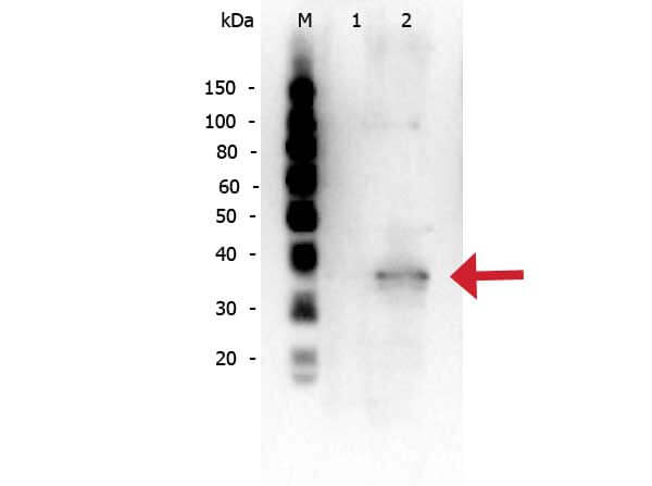
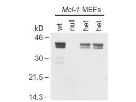
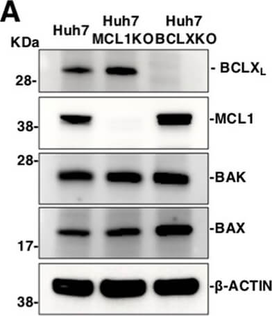
Western blot analysis using Rocklands anti-Mcl-1 antibody to detect Mcl-1 in MEF cell lysates (20 µg per lane). Anti-Mcl-1 was used at a dilution of 1:10,000 followed by reaction with HRP Anti-Rabbit IgG [H&L] MX10 (GOAT) used at a 1:2,000 dilution. Western Lightning Chemiluminescence Reagent Plus from Perkin Elmer was used for detection. The exposure time was exactly 30 seconds. This antibody detects 35 kDa mouse Mcl-1. Communicated with permission by J. Opferman and S. Korsmeyer. See Opferman et al (2003) for additional details. |
|
|
|
|
|
|
|
|
|
|
|
|
|
|
|
|
|
|
|
|
|
|
|
|
|
|
|
|
|
Product Guarantee and Expert Support
