Monoclonal Antibody to Human PD-L1 (Clone: ABM4E54)
Biozol Catalog Number:
ABI-10-7562
Supplier Catalog Number:
10-7562
Alternative Catalog Number:
ABI-10-7562-100UG
Manufacturer:
Abeomics
Host:
Mouse
Category:
Antikörper
Application:
FACS, IHC, WB
Species Reactivity:
Human
Immunogen:
A partial length recombinant PDL1 protein (amino acids 18-227) was used as the immunogen for this antibody.
Alternative Names:
CD274||B7H1||PDCD1L1||PDCD1LG1||PDL1
PD-L1 (CD274/B7-H1) is a critical membrane-bound costimulatory molecule belonging to the B7 superfamily that inhibits immune responses through its receptor, PD-1. PD-L1 plays a key role in the pathogenesis of inflammatory diseases (programmed death 1). It is widely expressed in the mononuclear phagocyte system (MPS), may co-stimulate T cells, and regulates inflammatory responses. PD-L1 exerts inflammation regulatory functions via a negative co-stimulatory effect on T cell functions to inhibit cytokine secretion, facilitate apoptosis of activated T cells and induce T cell anergy. Aberrant expression and dysregulation of CD274/PD-L1 have been reported during bacterial infection, inflammation, and in numerous autoimmune diseases.
Western blot analysis: 2-4 µg/ml, Immunohistochemical analysis-5-10 µg/ml, FACS: 1-2 µg/ml
Figure-2: Immunohistochemical analysis of PD-L1 in Hodgkins Lymphoma tissue using PD-L1 antibody (Clone: ABM4E54) at 5 µg/ml..
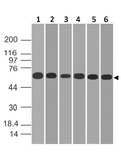
Figure-4: Western blot analysis of PD-L1. Anti-PD-L1 antibody (Clone: ABM4E54) was tested at 2 µg/ml on (1) A549, (2) MCF-7, (3) 293, (4) HCT-116, (5) Saos2 and (6) Hela lysates.
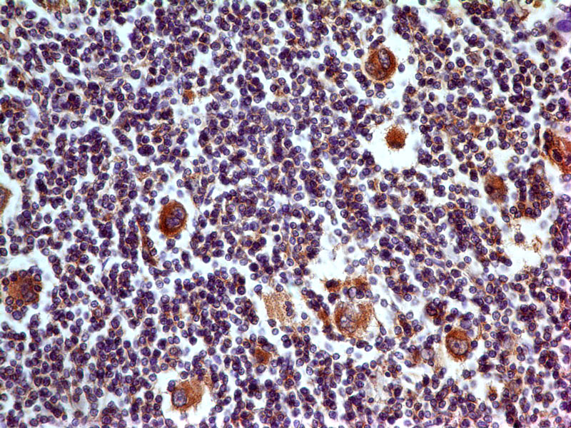
Figure-5: Immunohistochemical analysis of PD-L1 in Human Tonsil tissue using PD-L1 antibody (Clone: ABM4E54) at 5 µg/ml.
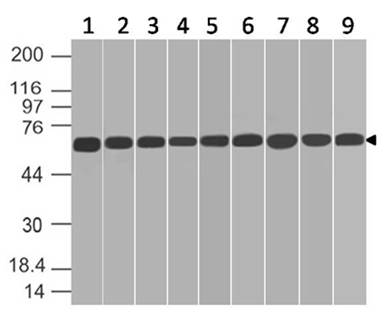
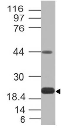
Figure-6: Western blot analysis of PD-L1. Anti-PD-L1 antibody (Clone: ABM4E54) was tested at 0.5 µg/ml on (1) HepG2, (2) SKBR3, (3) A431, (4) THP1, (5) NCCIT, (6) PC3, (7) PANC-1, (8) U87 and (9) KATO-111 lysates.
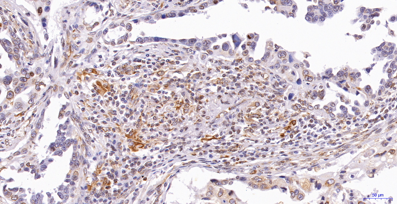
Figure-7: Immunohistochemical analysis of PD-L1 in Human Lung Cancer tissue using PD-L1 antibody (Clone: ABM4E54).
Figure-8: Western blot analysis of PD-L1. Anti-PD-L1 antibody (Clone: ABM4E54) was tested at 2 µg/ml on h Spleen lysate.
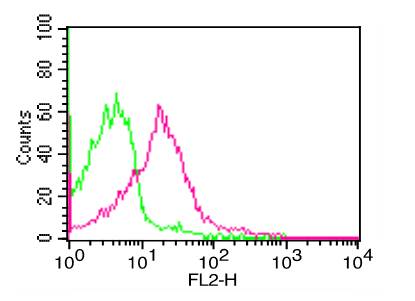
Figure-1: Cell Surface FLOW analysis of PD-L1 in PHA treated human PBMC using 1 µg of PD-L1 antibody (Clone: ABM4E54). Green represents isotype control, red represents anti-PD-L1 antibody. Goat anti-mouse PE conjugate was used as secondary antibody.
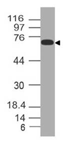
Figure-3: Western blot analysis of PD-L1. Anti-PD-L1 antibody (Clone: ABM4E54) was tested at 0.5 µg/ml on Recombinat lysates.
* VAT and and shipping costs not included. Errors and price changes excepted