This product is designed for immunofluorescence microscopy, fluorescence based plate assays (FLISA) and fluorescent western blotting. This product is also suitable for multiplex analysis, including multicolor imaging, utilizing various commercial platfor
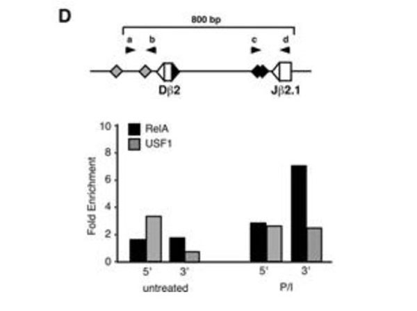
D, Protein-DNA complexes were immunoprecipitated overnight (4C) from 106cell equivalents sheared chromatin using protein G magnetic beads and antibodies specific for p65 RelA or upstream stimulatory factor 1 (USF1), or with normal rabbit IgG (p/n 110-4102). Chromatin IP analyses of NF-kappaB RelA and USF1 association with the 5' (primers aandb) and 3' Dbeta2 promoter sequences (primers candd) of endogenous P5424Tcrbbefore and after PMA/ionomycin treatment. Relative positions of the two NF-kappaB (♦) and predicted USF1 (gray diamonds) binding sites are indicated on the schematic diagram of the Dbeta2 region. Changes in the average Ct values of triplicate amplifications for specific Ab-bound samples relative to input controls were normalized to total serum IgG and transformed to generate fold enrichment over the isotype control. The results are representative of two separate experiments. FIGURE 6. PMID: 18292546.
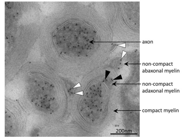
Immuno-electron micrograph detecting acetylated alpha-tubulin in axons and non-compact myelin. Acetylated microtubules were abundant in axons of mouse optic nerve as indicated by immuno-gold particles (black dots). In the adaxonal and abaxonal non-compact myelin compartment, acetylated microtubules were also present. Black and white arrow-heads point to corresponding immuno-gold particles in adaxonal and abaxonal non-compact myelin, respectively. On compact myelin, no immuno-gold was observed. Immunogold labeling of cryosections was performed on optic nerves of postnatal day 75. Antibody was specific for acetylated alpha-tubulin (1:1000) and was detected by incubation with rabbit anti-mouse IgG secondary antiserum (1:200 p/n 110-4102) which was visualized with protein A-gold (10 nm). Figure 4. PMID: 28248254.
* Mehrwertsteuer und Versandkosten nicht enthalten. Irrtümer und Preisänderungen vorbehalten