This product is designed for immunofluorescence microscopy, fluorescence based plate assays (FLISA) and fluorescent western blotting. This product is also suitable for multiplex analysis, including multicolor imaging, utilizing various commercial platfor
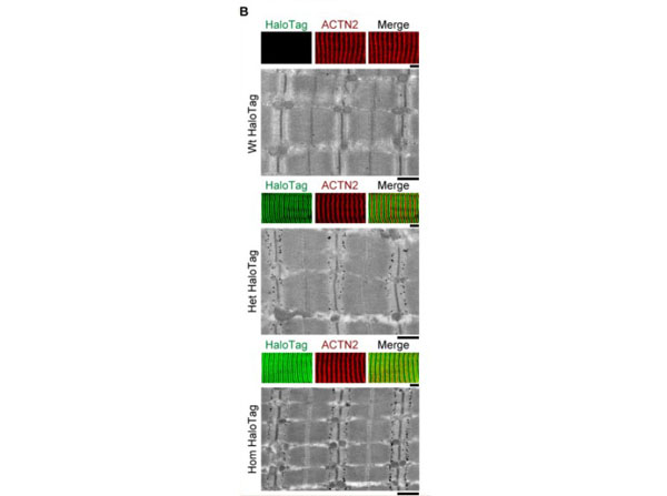
Mutant titin expression in skeletal muscles of different genotypes. (B) Correlative immunofluorescence (IF) and immunogold electron microscopy of wild-type (Wt), heterozygous (Het), and homozygous (Hom) psoas muscle. Representative IF images of fibers (colored panels) labeled with HaloTag antibody (green) and counterstained for alpha-actinin (ACTN2, red), and immunoelectron micrograph showing HaloTaglabeling. Scale bars, 5 µm (IF), 1 µm (IEM). Figure 1. PMID: 33357376.
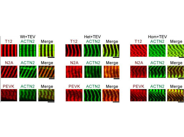
Immunofluorescence (IF) micrographs of skeletal fibers labeled with different antibodies to I-band titin (T12, N2A, and PEVK, red IF staining), and Z-disk marker alpha-actinin (ACTN2, green IF staining). Shown are examples of Wt, Het, and Hom fibers after TEV-protease treatment, held passively for 30 min at a stretched length. Scale bars, 5 µm. Figure 5S1. PMID: 33357376.
* Mehrwertsteuer und Versandkosten nicht enthalten. Irrtümer und Preisänderungen vorbehalten