0.02 M Potassium Phosphate, 0.15 M Sodium Chloride, pH 7.2
Formulierung:
Lyophilized
Target-Kategorie:
Mouse
Antibody Type:
Secondary Antibody
Application Verdünnung:
FLISA: >1:20,000, IF Microscopy: >1:5,000, WB: >1:10,000
Anwendungsbeschreibung:
Anti-Mouse IgG ATTO425 Antibody has been tested by dot blot and western blot and is designed for STED microscopy, FRET, immunofluorescence microscopy, fluorescence based plate assays (FLISA) and fluorescent western blotting. Rabbit anti-mouse IgG antibod
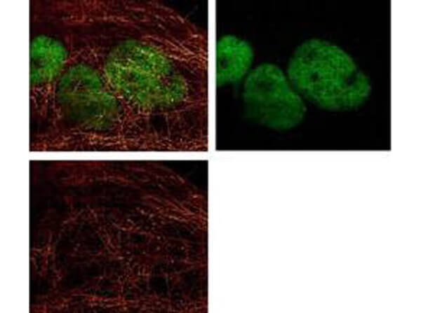
ATTO 425 conjugated anti-Mouse IgG was used to demonstrate 2 color STED immunofluorescence microscopy. Methanol fixed A431 cells were blocked with normal goat serum. The cells were then probed with 0.4 µg/mL final concentration of anti-a-tubulin and detected with 0.2 µg/mL ATTO 425 conjugated anti-MOUSE IgG [GOAT] (610-151-121) secondary antibody (colored RED). Also shown in this 2-color STED image is Rocklands Anti-HDAC-1 [RABBIT] (p/n 600-401-879) detected with DyLight(TM)488 conjugated Anti-RABBIT IgG [GOAT] secondary antibody (colored GREEN). Image courtesy of Myriam Gastard, Leica Microsystems, USA.
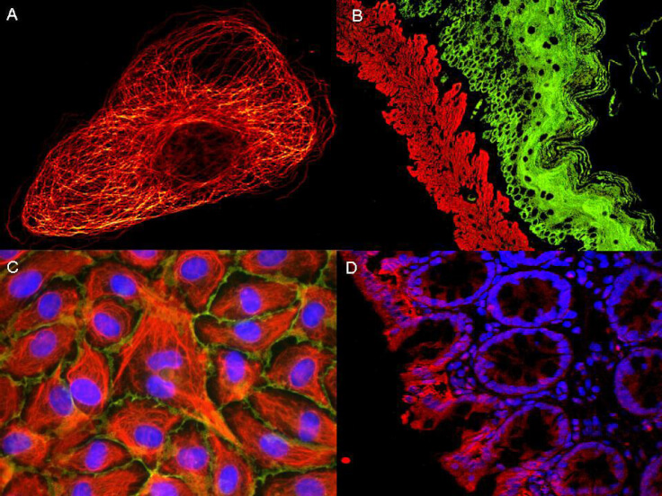
ATTO dyes can be used for multicolor immunofluorescent detection with low background and high signal. Examples shown are: A. Tubulin in PtK2- male Rat Kangaroo Kidney Epithelial Cells was detected using ATTO 532 labeled secondary antibody. B. Muscle alpha-actin was stained with a mouse primary antibody and ATTO 488 anti-mouse IgG (green) while Cytokeratin was stained with polyclonal rabbit anti-cytokeratin and ATTO 647N anti-rabbit IgG (red). C. HUVEC (Human umbilical vein endothelial cells were stained with anti- Vimentin-ATTO 532 (green), anti-E-Cadherin-ATTO 655 (red) and DAPI (blue). D. Rat colon sections were stained with Anti-Aquaporin 3-ATTO 594 antibody. Hoechst 33342 (blue) is used as counterstain. Images provided courtesy of Dr. Jörg Reichwein, ATTO-TEC GmbH
* Mehrwertsteuer und Versandkosten nicht enthalten. Irrtümer und Preisänderungen vorbehalten