
MMP14/MT1-MMP Rabbit mAb, Unconjugated, Monoclonal
Artikelnummer:
ABB-A0067
- Bilder (9)


| Artikelname: | MMP14/MT1-MMP Rabbit mAb, Unconjugated, Monoclonal |
| Artikelnummer: | ABB-A0067 |
| Hersteller Artikelnummer: | A0067 |
| Alternativnummer: | ABB-A0067-100UL,ABB-A0067-20UL |
| Hersteller: | ABclonal |
| Wirt: | Rabbit |
| Kategorie: | Antikörper |
| Applikation: | ELISA, FC, IHC-P, WB |
| Spezies Reaktivität: | Human |
| Immunogen: | Synthetic peptide. This information is considered to be commercially sensitive. |
| Konjugation: | Unconjugated |
| Alternative Synonym: | MMP-14, MMP-X1, MT-MMP, MT1MMP, MTMMP1, WNCHRS, MT1-MMP, MT-MMP 1, MMP14/MT1-MMP |
| Proteins of the matrix metalloproteinase (MMP) family are involved in the breakdown of extracellular matrix in normal physiological processes, such as embryonic development, reproduction, and tissue remodeling, as well as in disease processes, such as arthritis and metastasis. Most MMPs are secreted as inactive proproteins which are activated when cleaved by extracellular proteinases. However, the protein encoded by this gene is a member of the membrane-type MMP (MT-MMP) subfamily, each member of this subfamily contains a potential transmembrane domain suggesting that these proteins are expressed at the cell surface rather than secreted. This protein activates MMP2 protein, and this activity may be involved in tumor invasion. |
| Application Verdünnung: | WB,1:500 - 1:2000|IHC-P, 1:50 - 1:200|FC, 1:100 - 1:500|ELISA,Recommended starting concentration is 1 µg/mL. Please optimize the concentration based on your specific assay requirements. |
| Anwendungsbeschreibung: | Cross-Reactivity: Human,Mouse,Rat. ResearchArea: Cancer,Tumor biomarkers,Invasion and Metastasis,Signal Transduction,Cell Biology Developmental Biology,Cytoskeleton,Extracellular Matrix,Ubiquitin,Endocrine Metabolism,Cardiovascular,Angiogenesis,Hypoxia. Shipping: Ice Bag |
|
|
Western blot analysis of various lysates using MMP14/MMP14/MT1-MMP Rabbit mAb (A0067) at 1:1000 dilution. Secondary antibody: HRP-conjugated Goat anti-Rabbit IgG (H+L) (AS014) at 1:10000 dilution. Lysates/proteins: 25µg per lane. Blocking buffer: 3% nonfat dry milk in TBST. Detection: ECL Basic Kit (RM00020). Exposure time: 90s. |
|
|
Western blot analysis of various lysates using MMP14/MMP14/MT1-MMP Rabbit mAb (A0067) at 1:1000 dilution. Secondary antibody: HRP-conjugated Goat anti-Rabbit IgG (H+L) (AS014) at 1:10000 dilution. Lysates/proteins: 25µg per lane. Blocking buffer: 3% nonfat dry milk in TBST. Detection: ECL Basic Kit (RM00020). Exposure time: 10s. |
|
|
Immunohistochemistry analysis of paraffin-embedded Human liver tissue using MMP14/MT1-MMP Rabbit mAb (A0067) at a dilution of 1:200 (40x lens). High pressure antigen retrieval performed with 0.01M Citrate buffer (pH 6.0) prior to IHC staining. |
|
|
Immunohistochemistry analysis of paraffin-embedded Human spleen tissue using MMP14/MT1-MMP Rabbit mAb (A0067) at a dilution of 1:200 (40x lens). High pressure antigen retrieval performed with 0.01M Citrate buffer (pH 6.0) prior to IHC staining. |
|
|
Immunohistochemistry analysis of paraffin-embedded Mouse kidney tissue using MMP14/MT1-MMP Rabbit mAb (A0067) at a dilution of 1:200 (40x lens). High pressure antigen retrieval performed with 0.01M Citrate buffer (pH 6.0) prior to IHC staining. |
|
|
Flow cytometry: 1X10 6 MCF7 cells (negative control,left) and HT-1080 (right) cells were intracellularly-stained with MMP14/MT1-MMP Rabbit mAb (A0067,2 µg/mL,orange line) or ABflo 488 Rabbit IgG isotype control (AC042,2 µg/mL,blue line), followed by Alexa Fluor 488 conjugated goat anti-rabbit pAb staining. Non-fluorescently stained cells were used as blank control (red line). |
|
|
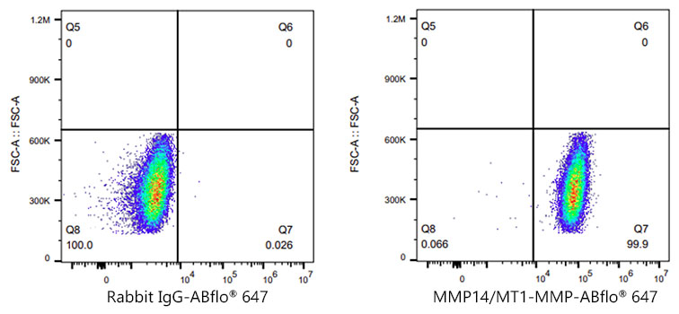
Flow cytometry: 1X10 6 MCF7 cells (negative control,left) and HT-1080 (right) cells were intracellularly-stained with MMP14/MT1-MMP Rabbit mAb (A0067,2 µg/mL,orange line) or ABflo 647 Rabbit IgG isotype control (AC042,2 µg/mL,blue line), followed by Alexa Fluor 647 conjugated goat anti-rabbit pAb staining. Non-fluorescently stained cells were used as blank control (red line). |
|
|
Flow cytometry: 1X10 6 MCF7 cells (negative control,left) and HT-1080 (right) cells were intracellularly-stained with MMP14/MT1-MMP Rabbit mAb (A0067,2 µg/mL,orange line) or ABflo 647 Rabbit IgG isotype control (AC042,2 µg/mL,blue line), followed by Alexa Fluor 647 conjugated goat anti-rabbit pAb staining. Non-fluorescently stained cells were used as blank control (red line). |
Produktgarantie und fachkundiger Support
