
PRP19 Rabbit pAb, Unconjugated, Polyclonal
Artikelnummer:
ABB-A12590
- Bilder (9)


| Artikelname: | PRP19 Rabbit pAb, Unconjugated, Polyclonal |
| Artikelnummer: | ABB-A12590 |
| Hersteller Artikelnummer: | A12590 |
| Alternativnummer: | ABB-A12590-100UL,ABB-A12590-20UL,ABB-A12590-1000UL,ABB-A12590-500UL |
| Hersteller: | ABclonal |
| Wirt: | Rabbit |
| Kategorie: | Antikörper |
| Applikation: | ELISA, IF, IHC-P, IP, WB |
| Spezies Reaktivität: | Human |
| Immunogen: | Recombinant protein (or fragment).This information is considered to be commercially sensitive. |
| Konjugation: | Unconjugated |
| Alternative Synonym: | PSO4, SNEV, PRP19, UBOX4, hPSO4, NMP200 |
| Enables identical protein binding activity and ubiquitin-ubiquitin ligase activity. Involved in several processes, including DNA damage checkpoint signaling, cellular protein metabolic process, and mRNA splicing, via spliceosome. Acts upstream of or within protein polyubiquitination. Located in cytoplasm, nuclear speck, and site of double-strand break. Part of Prp19 complex and U2-type catalytic step 2 spliceosome. Colocalizes with DNA replication factor A complex. |
| Klonalität: | Polyclonal |
| Molekulargewicht: | 55kDa |
| NCBI: | 27339 |
| UniProt: | Q9UMS4 |
| Reinheit: | Affinity purification |
| Sequenz: | AREALATLKPQAGLIVPQAVPSSQPSVVGAGEPMDLGELVGMTPEIIQKLQDKATVLTTERKKRGKTVPEELVKPEELSKYRQVASHVGLHSASIPGILALDLCPSDTNKILTGGADKNVVVFDKSSEQILATLKGHTKKVTSVVFHPSQDLVFSASPDATIRIWSVPNASCVQVVRAHESAVTGLSLHATGDYLLSSSDDQYWAFSDIQTGRVLTKVTDETSGCSLTCAQFHPDGLIFGTGTMDSQIKIWDLKE |
| Target-Kategorie: | PRPF19 |
| Antibody Type: | Primary Antibody |
| Application Verdünnung: | WB,1:500 - 1:2000|IHC-P,1:50 - 1:200|IF/ICC,1:50 - 1:200|IP,0.5µg-4µg antibody for 200µg-400µg extracts of whole cells|ELISA,Recommended starting concentration is 1 µg/mL. Please optimize the concentration based on your specific assay requirements. |
| Anwendungsbeschreibung: | Cross-Reactivity: Human,Mouse,Rat. ResearchArea: Epigenetics Nuclear Signaling,DNA Damage Repair,Cell Biology Developmental Biology,Ubiquitin,Ubiquitin-Proteasome Signaling Pathway. Shipping: Ice Bag |
|
|
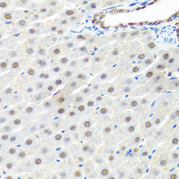
Immunohistochemistry analysis of paraffin-embedded Rat liver using PRP19 Rabbit pAb (A12590) at dilution of 1:100 (40x lens). Microwave antigen retrieval performed with 0.01M PBS Buffer (pH 7.2) prior to IHC staining. |
|
|
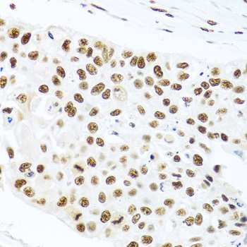
Immunohistochemistry analysis of paraffin-embedded Human lung cancer using PRP19 Rabbit pAb (A12590) at dilution of 1:100 (40x lens). Microwave antigen retrieval performed with 0.01M PBS Buffer (pH 7.2) prior to IHC staining. |
|
|
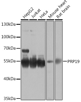
Western blot analysis of various lysates using PRP19 Rabbit pAb (A12590) at 1:1000 dilution. Secondary antibody: HRP-conjugated Goat anti-Rabbit IgG (H+L) (AS014) at 1:10000 dilution. Lysates/proteins: 25µg per lane. Blocking buffer: 3% nonfat dry milk in TBST. Detection: ECL Basic Kit (RM00020). Exposure time: 30s. |
|
|
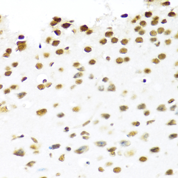
Immunohistochemistry analysis of paraffin-embedded Human stomach using PRP19 Rabbit pAb (A12590) at dilution of 1:100 (40x lens). Microwave antigen retrieval performed with 0.01M PBS Buffer (pH 7.2) prior to IHC staining. |
|
|
Immunohistochemistry analysis of paraffin-embedded Mouse brain using PRP19 Rabbit pAb (A12590) at dilution of 1:100 (40x lens). Microwave antigen retrieval performed with 0.01M PBS Buffer (pH 7.2) prior to IHC staining. |
|
|
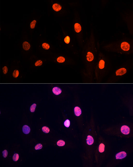
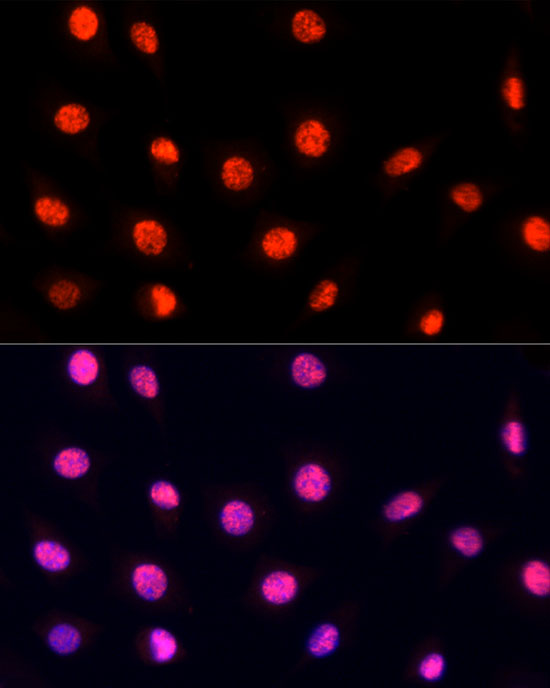
Immunofluorescence analysis of H9C2 cells using PRP19 Rabbit pAb (A12590) at dilution of 1:100 (40x lens). Secondary antibody: Cy3-conjugated Goat anti-Rabbit IgG (H+L) (AS007) at 1:500 dilution. Blue: DAPI for nuclear staining. |
|
|
Immunofluorescence analysis of L929 cells using PRP19 Rabbit pAb (A12590) at dilution of 1:100 (40x lens). Secondary antibody: Cy3-conjugated Goat anti-Rabbit IgG (H+L) (AS007) at 1:500 dilution. Blue: DAPI for nuclear staining. |
|
|
Immunofluorescence analysis of U2OS cells using PRP19 Rabbit pAb (A12590) at dilution of 1:100 (40x lens). Secondary antibody: Cy3-conjugated Goat anti-Rabbit IgG (H+L) (AS007) at 1:500 dilution. Blue: DAPI for nuclear staining. |
|
|
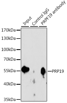
Immunoprecipitation analysis of 200 µg extracts of Jurkat cells, using 3 µg PRP19 antibody (A12590). Western blot was performed from the immunoprecipitate using PRP19 antibody (A12590) at a dilution of 1:1000. |
Produktgarantie und fachkundiger Support
