
Monoamine Oxidase B (MAOB) Rabbit pAb, Unconjugated, Polyclonal
Artikelnummer:
ABB-A1568
- Bilder (9)


| Artikelname: | Monoamine Oxidase B (MAOB) Rabbit pAb, Unconjugated, Polyclonal |
| Artikelnummer: | ABB-A1568 |
| Hersteller Artikelnummer: | A1568 |
| Alternativnummer: | ABB-A1568-100UL,ABB-A1568-20UL,ABB-A1568-1000UL,ABB-A1568-500UL |
| Hersteller: | ABclonal |
| Wirt: | Rabbit |
| Kategorie: | Antikörper |
| Applikation: | ELISA, IF, IHC-P, WB |
| Spezies Reaktivität: | Human |
| Immunogen: | Recombinant protein (or fragment).This information is considered to be commercially sensitive. |
| Konjugation: | Unconjugated |
| Alternative Synonym: | MAOB, Monoamine Oxidase B (MAOB) |
| The protein encoded by this gene belongs to the flavin monoamine oxidase family. It is a enzyme located in the mitochondrial outer membrane. It catalyzes the oxidative deamination of biogenic and xenobiotic amines and plays an important role in the metabolism of neuroactive and vasoactive amines in the central nervous sysytem and peripheral tissues. This protein preferentially degrades benzylamine and phenylethylamine. |
| Application Verdünnung: | WB,1:500 - 1:1000|IHC-P,1:50 - 1:200|IF/ICC,1:50 - 1:200|ELISA,Recommended starting concentration is 1 µg/mL. Please optimize the concentration based on your specific assay requirements. |
| Anwendungsbeschreibung: | Cross-Reactivity: Human,Mouse,Rat. ResearchArea: Cancer,Signal Transduction,Endocrine Metabolism,Amino acid metabolism,Drug metabolism,Neuroscience,Neurodegenerative Diseases,Dopamine Signaling in Parkinsons Disease. Shipping: Ice Bag |
|
|
Immunohistochemistry analysis of paraffin-embedded Rat liver using Monoamine Oxidase B (MAOB) Rabbit pAb (A1568) at dilution of 1:100 (40x lens). High pressure antigen retrieval performed with 0.01M Citrate buffer (pH 6.0) prior to IHC staining. |
|
|
Immunohistochemistry analysis of paraffin-embedded Rat heart using Monoamine Oxidase B (MAOB) Rabbit pAb (A1568) at dilution of 1:100 (40x lens). High pressure antigen retrieval performed with 0.01M Citrate buffer (pH 6.0) prior to IHC staining. |
|
|
Western blot analysis of lysates from HeLa cells, using Monoamine Oxidase B (MAOB) Rabbit pAb (A1568) at 1:1000 dilution. Secondary antibody: HRP-conjugated Goat anti-Rabbit IgG (H+L) (AS014) at 1:10000 dilution. Lysates/proteins: 25µg per lane. Blocking buffer: 3% nonfat dry milk in TBST. Detection: ECL Basic Kit (RM00020). Exposure time: 1s. |
|
|
Immunohistochemistry analysis of paraffin-embedded Human liver using Monoamine Oxidase B (MAOB) Rabbit pAb (A1568) at dilution of 1:100 (40x lens). High pressure antigen retrieval performed with 0.01M Citrate buffer (pH 6.0) prior to IHC staining. |
|
|
Immunohistochemistry analysis of paraffin-embedded Human liver cancer using Monoamine Oxidase B (MAOB) Rabbit pAb (A1568) at dilution of 1:100 (40x lens). High pressure antigen retrieval performed with 0.01M Citrate buffer (pH 6.0) prior to IHC staining. |
|
|
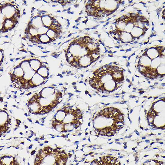
Immunohistochemistry analysis of paraffin-embedded Human colon using Monoamine Oxidase B (MAOB) Rabbit pAb (A1568) at dilution of 1:100 (40x lens). High pressure antigen retrieval performed with 0.01M Citrate buffer (pH 6.0) prior to IHC staining. |
|
|
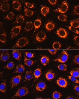
Immunofluorescence analysis of L929 cells using Monoamine Oxidase B (Monoamine Oxidase B (MAOB)) Rabbit pAb (A1568) at dilution of 1:100. Secondary antibody: Cy3-conjugated Goat anti-Rabbit IgG (H+L) (AS007) at 1:500 dilution. Blue: DAPI for nuclear staining. |
|
|
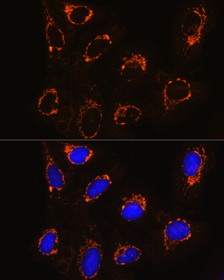
Immunofluorescence analysis of U2OS cells using Monoamine Oxidase B (Monoamine Oxidase B (MAOB)) Rabbit pAb (A1568) at dilution of 1:100. Secondary antibody: Cy3-conjugated Goat anti-Rabbit IgG (H+L) (AS007) at 1:500 dilution. Blue: DAPI for nuclear staining. |
|
|
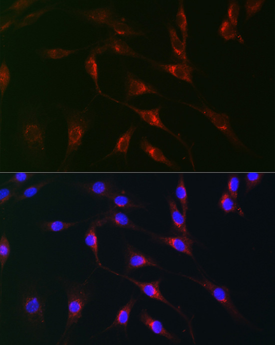
Immunofluorescence analysis of NIH/3T3 cells using Monoamine Oxidase B (MAOB) Rabbit pAb (A1568) at dilution of 1:100 (40x lens). Secondary antibody: Cy3-conjugated Goat anti-Rabbit IgG (H+L) (AS007) at 1:500 dilution. Blue: DAPI for nuclear staining. |
Produktgarantie und fachkundiger Support
