
Annexin A2 Rabbit pAb, Unconjugated, Polyclonal
Artikelnummer:
ABB-A1572
- Bilder (9)


| Artikelname: | Annexin A2 Rabbit pAb, Unconjugated, Polyclonal |
| Artikelnummer: | ABB-A1572 |
| Hersteller Artikelnummer: | A1572 |
| Alternativnummer: | ABB-A1572-1000UL,ABB-A1572-100UL,ABB-A1572-500UL,ABB-A1572-20UL |
| Hersteller: | ABclonal |
| Wirt: | Rabbit |
| Kategorie: | Antikörper |
| Applikation: | ELISA, IF, IHC-P, WB |
| Spezies Reaktivität: | Human |
| Immunogen: | Recombinant protein (or fragment).This information is considered to be commercially sensitive. |
| Konjugation: | Unconjugated |
| Alternative Synonym: | P36, ANX2, LIP2, LPC2, CAL1H, LPC2D, ANX2L4, PAP-IV, HEL-S-270, Annexin A2 |
| This gene encodes a member of the annexin family. Members of this calcium-dependent phospholipid-binding protein family play a role in the regulation of cellular growth and in signal transduction pathways. This protein functions as an autocrine factor which heightens osteoclast formation and bone resorption. This gene has three pseudogenes located on chromosomes 4, 9 and 10, respectively. Multiple alternatively spliced transcript variants encoding different isoforms have been found for this gene. Annexin A2 expression has been found to correlate with resistance to treatment against various cancer forms. |
| Klonalität: | Polyclonal |
| Molekulargewicht: | 39kDa |
| NCBI: | 302 |
| UniProt: | P07355 |
| Reinheit: | Affinity purification |
| Sequenz: | MSTVHEILCKLSLEGDHSTPPSAYGSVKAYTNFDAERDALNIETAIKTKGVDEVTIVNILTNRSNAQRQDIAFAYQRRTKKELASALKSALSGHLETVILGLLKTPAQYDASELKASMKGLGTDEDSLIEIICSRTNQELQEINRVYKEMYKTDLEKDIISDTSGDFRKLMVALAKGRRAEDGSVIDYELIDQDARDLYDAGVKRKGTDVPKWISIMTERSVPHLQKVFDRYKSYSPYDMLESIRKEVKGDLENA |
| Target-Kategorie: | ANXA2 |
| Application Verdünnung: | WB,1:500 - 1:1000|IHC-P,1:50 - 1:200|IF/ICC,1:50 - 1:200|ELISA,Recommended starting concentration is 1 µg/mL. Please optimize the concentration based on your specific assay requirements. |
| Anwendungsbeschreibung: | Cross-Reactivity: Human,Mouse,Rat. ResearchArea: Epigenetics Nuclear Signaling,RNA Binding,Signal Transduction,Cell Biology Developmental Biology,Cell Adhesion,Cytoskeleton,Neuroscience,Calcium Signaling. Shipping: Ice Bag |
|
|
Western blot analysis of various lysates using Annexin A2 Rabbit pAb (A1572) at 1:500 dilution. Secondary antibody: HRP-conjugated Goat anti-Rabbit IgG (H+L) (AS014) at 1:10000 dilution. Lysates/proteins: 25µg per lane. Blocking buffer: 3% nonfat dry milk in TBST. Detection: ECL Basic Kit (RM00020). Exposure time: 60s. |
|
|
Immunohistochemistry analysis of paraffin-embedded Human liver cancer using Annexin A2 Rabbit pAb (A1572) at dilution of 1:20 (40x lens). High pressure antigen retrieval performed with 0.01M Citrate buffer (pH 6.0) prior to IHC staining. |
|
|
Western blot analysis of various lysates using Annexin A2 Rabbit pAb (A1572) at 1:500 dilution. Secondary antibody: HRP-conjugated Goat anti-Rabbit IgG (H+L) (AS014) at 1:10000 dilution. Lysates/proteins: 25µg per lane. Blocking buffer: 3% nonfat dry milk in TBST. Detection: ECL Basic Kit (RM00020). Exposure time: 10s. |
|
|
Immunohistochemistry analysis of paraffin-embedded Human liver using Annexin A2 Rabbit pAb (A1572) at dilution of 1:20 (40x lens). High pressure antigen retrieval performed with 0.01M Citrate buffer (pH 6.0) prior to IHC staining. |
|
|
Immunohistochemistry analysis of paraffin-embedded Mouse kidney using Annexin A2 Rabbit pAb (A1572) at dilution of 1:20 (40x lens). High pressure antigen retrieval performed with 0.01M Citrate buffer (pH 6.0) prior to IHC staining. |
|
|
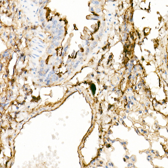
Immunohistochemistry analysis of paraffin-embedded Mouse lung using Annexin A2 Rabbit pAb (A1572) at dilution of 1:20 (40x lens). High pressure antigen retrieval performed with 0.01M Citrate buffer (pH 6.0) prior to IHC staining. |
|
|
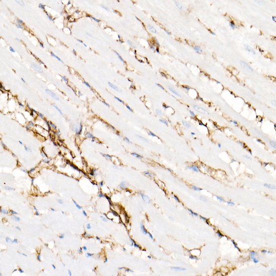
Immunohistochemistry analysis of paraffin-embedded Rat heart using Annexin A2 Rabbit pAb (A1572) at dilution of 1:20 (40x lens). High pressure antigen retrieval performed with 0.01M Citrate buffer (pH 6.0) prior to IHC staining. |
|
|
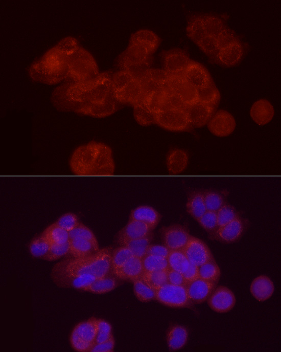
Immunofluorescence analysis of MCF7 cells using Annexin A2 Rabbit pAb (A1572) at dilution of 1:20 (40x lens). Secondary antibody: Cy3-conjugated Goat anti-Rabbit IgG (H+L) (AS007) at 1:500 dilution. Blue: DAPI for nuclear staining. |
|
|
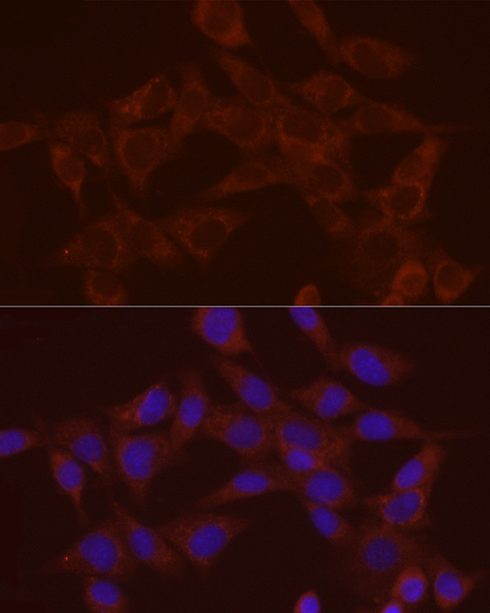
Immunofluorescence analysis of NIH/3T3 cells using Annexin A2 Rabbit pAb (A1572) at dilution of 1:20 (40x lens). Secondary antibody: Cy3-conjugated Goat anti-Rabbit IgG (H+L) (AS007) at 1:500 dilution. Blue: DAPI for nuclear staining. |
Produktgarantie und fachkundiger Support
