
[KO Validated] Integrin alpha 2 (ITGA2/CD49b) Rabbit mAb, Unconjugated, Monoclonal
Artikelnummer:
ABB-A19068
- Bilder (9)

| Artikelname: | [KO Validated] Integrin alpha 2 (ITGA2/CD49b) Rabbit mAb, Unconjugated, Monoclonal |
| Artikelnummer: | ABB-A19068 |
| Hersteller Artikelnummer: | A19068 |
| Alternativnummer: | ABB-A19068-20UL,ABB-A19068-100UL,ABB-A19068-1000UL,ABB-A19068-500UL |
| Hersteller: | ABclonal |
| Wirt: | Rabbit |
| Kategorie: | Antikörper |
| Applikation: | ELISA, IHC-P, WB |
| Spezies Reaktivität: | Human |
| Immunogen: | Synthetic peptide. This information is considered to be commercially sensitive. |
| Konjugation: | Unconjugated |
| Alternative Synonym: | BR, GPIa, CD49B, HPA-5, VLA-2, VLAA2, b) |
| This gene encodes the alpha subunit of a transmembrane receptor for collagens and related proteins. The encoded protein forms a heterodimer with a beta subunit and mediates the adhesion of platelets and other cell types to the extracellular matrix. Loss of the encoded protein is associated with bleeding disorder platelet-type 9. Antibodies against this protein are found in several immune disorders, including neonatal alloimmune thrombocytopenia. This gene is located adjacent to a related alpha subunit gene. Alternative splicing results in multiple transcript variants. |
| Klonalität: | Monoclonal |
| Klon-Bezeichnung: | [ARC0457] |
| Molekulargewicht: | 129kDa |
| NCBI: | 3673 |
| UniProt: | P17301 |
| Reinheit: | Affinity purification |
| Sequenz: | GTFASSTFQTVQLTAAAEINTYNPEIYVIEDNTVTIPLMIMKPDEKAEVPTGVIIGSIIAGILLLLALVAILWKLGFFKRKYEKMTKNPDEIDETTELSS |
| Target-Kategorie: | ITGA2 |
| Antibody Type: | Primary Antibody |
| Application Verdünnung: | WB,1:1000 - 1:6000|IHC-P,1:200 - 1:800|ELISA,Recommended starting concentration is 1 µg/mL. Please optimize the concentration based on your specific assay requirements. |
| Anwendungsbeschreibung: | Cross-Reactivity: Human,Mouse,Rat. ResearchArea: Cancer,Signal Transduction,PI3K-Akt Signaling Pathway,MAPK-Erk Signaling Pathway,Cell Biology Developmental Biology,Cell Adhesion,Cytoskeleton,Endocrine Metabolism,Immunology Inflammation,CDs. Shipping: Ice Bag |
|
|
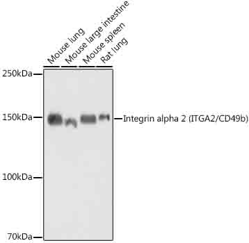
Western blot analysis of various lysates, using [KO Validated] Integrin alpha 2 (ITGA2/CD49b) Rabbit mAb (A19068) at 1:1000 dilution. Secondary antibody: HRP-conjugated Goat anti-Rabbit IgG (H+L) (AS014) at 1:10000 dilution. Lysates/proteins: 25µg per lane. Blocking buffer: 3% nonfat dry milk in TBST. Detection: ECL Basic Kit (RM00020). Exposure time: 10s. |
|
|
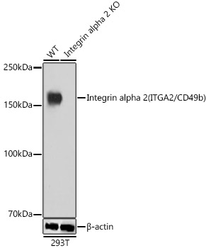
Western blot analysis of lysates from wild type(WT) and Integrin alpha 2 (ITGA2/CD49b) knockout (KO) 293T cells, using [KO Validated] Integrin alpha 2 (ITGA2/CD49b) Rabbit mAb (A19068) at 1:1000 dilution. Secondary antibody: HRP-conjugated Goat anti-Rabbit IgG (H+L) (AS014) at 1:10000 dilution. Lysates/proteins: 25µg per lane. Blocking buffer: 3% nonfat dry milk in TBST. Detection: ECL Basic Kit (RM00020). Exposure time: 1s. |
|
|
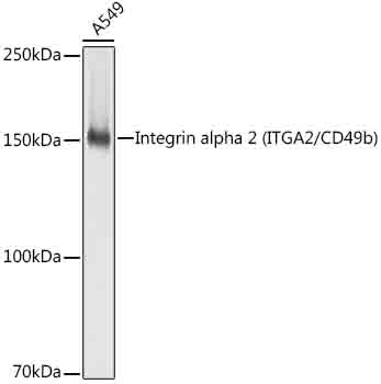
Western blot analysis of lysates from A549 cells, using [KO Validated] Integrin alpha 2 (ITGA2/CD49b) Rabbit mAb (A19068) at 1:1000 dilution. Secondary antibody: HRP-conjugated Goat anti-Rabbit IgG (H+L) (AS014) at 1:10000 dilution. Lysates/proteins: 25µg per lane. Blocking buffer: 3% nonfat dry milk in TBST. Detection: ECL Basic Kit (RM00020). Exposure time: 1s. |
|
|
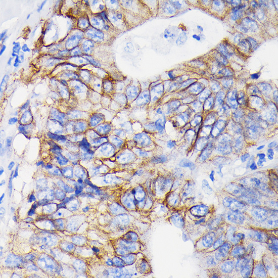
Immunohistochemistry analysis of paraffin-embedded Human colon carcinoma tissue using [KO Validated] Integrin alpha 2 (ITGA2/CD49b) Rabbit mAb (A19068) at a dilution of 1:500 (40x lens). High pressure antigen retrieval performed with 0.01M Tris-EDTA Buffer (pH 9.0) prior to IHC staining. |
|
|
Immunohistochemistry analysis of paraffin-embedded Human breast tissue using [KO Validated] Integrin alpha 2 (ITGA2/CD49b) Rabbit mAb (A19068) at a dilution of 1:500 (40x lens). High pressure antigen retrieval performed with 0.01M Tris-EDTA Buffer (pH 9.0) prior to IHC staining. |
|
|
Immunohistochemistry analysis of paraffin-embedded Human esophagus tissue using [KO Validated] Integrin alpha 2 (ITGA2/CD49b) Rabbit mAb (A19068) at a dilution of 1:500 (40x lens). High pressure antigen retrieval performed with 0.01M Tris-EDTA Buffer (pH 9.0) prior to IHC staining. |
|
|
Immunohistochemistry analysis of paraffin-embedded Human kidney tissue using [KO Validated] Integrin alpha 2 (ITGA2/CD49b) Rabbit mAb (A19068) at a dilution of 1:500 (40x lens). High pressure antigen retrieval performed with 0.01M Tris-EDTA Buffer (pH 9.0) prior to IHC staining. |
|
|
Immunohistochemistry analysis of paraffin-embedded Mouse spleen tissue using [KO Validated] Integrin alpha 2 (ITGA2/CD49b) Rabbit mAb (A19068) at a dilution of 1:500 (40x lens). High pressure antigen retrieval performed with 0.01M Tris-EDTA Buffer (pH 9.0) prior to IHC staining. |
|
|
Immunohistochemistry analysis of paraffin-embedded Rat colon tissue using [KO Validated] Integrin alpha 2 (ITGA2/CD49b) Rabbit mAb (A19068) at a dilution of 1:500 (40x lens). High pressure antigen retrieval performed with 0.01M Tris-EDTA Buffer (pH 9.0) prior to IHC staining. |
Produktgarantie und fachkundiger Support
