
VDAC1 Rabbit mAb, Unconjugated, Monoclonal
Artikelnummer:
ABB-A19707
- Bilder (9)


| Artikelname: | VDAC1 Rabbit mAb, Unconjugated, Monoclonal |
| Artikelnummer: | ABB-A19707 |
| Hersteller Artikelnummer: | A19707 |
| Alternativnummer: | ABB-A19707-100UL,ABB-A19707-20UL,ABB-A19707-1000UL,ABB-A19707-500UL |
| Hersteller: | ABclonal |
| Wirt: | Rabbit |
| Kategorie: | Antikörper |
| Applikation: | ELISA, IF, IHC-P, WB |
| Spezies Reaktivität: | Human |
| Immunogen: | A synthetic peptide corresponding to a sequence within amino acids 1-100 of human VDAC1 (P21796). |
| Konjugation: | Unconjugated |
| Alternative Synonym: | PORIN, VDAC-1, VDAC1 |
| This gene encodes a voltage-dependent anion channel protein that is a major component of the outer mitochondrial membrane. The encoded protein facilitates the exchange of metabolites and ions across the outer mitochondrial membrane and may regulate mitochondrial functions. This protein also forms channels in the plasma membrane and may be involved in transmembrane electron transport. Alternate splicing results in multiple transcript variants. Multiple pseudogenes of this gene are found on chromosomes 1, 2 3, 6, 9, 12, X and Y. |
| Application Verdünnung: | WB,1:5000 - 1:20000|IHC-P,1:1000 - 1:4000|IF/ICC,1:200 - 1:500|ELISA,Recommended starting concentration is 1 µg/mL. Please optimize the concentration based on your specific assay requirements |
| Anwendungsbeschreibung: | Cross-Reactivity: Human,Mouse,Rat. ResearchArea: Cancer,Signal Transduction,Endocrine Metabolism,Mitochondrial metabolism,Mitochondrial markers,Warburg Effect,Neuroscience,Neurodegenerative Diseases. Shipping: Ice Bag |
|
|
Confocal imaging of C6 cells using VDAC1 Rabbit mAb (A19707, dilution 1:200) followed by a further incubation with Cy3 Goat Anti-Rabbit IgG (H+L) (AS007, dilution 1:500) (Red). The cells were counterstained with alpha-Tubulin Mouse mAb (AC012, dilution 1:400) followed by incubation with ABflo 488-conjugated Goat Anti-Mouse IgG (H+L) Ab (AS076, dilution 1:500) (Green). DAPI was used for nuclear staining (Blue). Objective: 100x. |
|
|
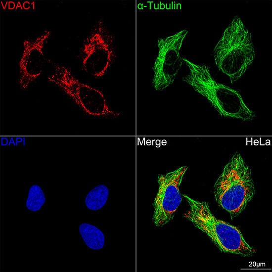
Confocal imaging of HeLa cells using VDAC1 Rabbit mAb (A19707, dilution 1:200) followed by a further incubation with Cy3 Goat Anti-Rabbit IgG (H+L) (AS007, dilution 1:500) (Red). The cells were counterstained with alpha-Tubulin Mouse mAb (AC012, dilution 1:400) followed by incubation with ABflo 488-conjugated Goat Anti-Mouse IgG (H+L) Ab (AS076, dilution 1:500) (Green). DAPI was used for nuclear staining (Blue). Objective: 100x. |
|
|
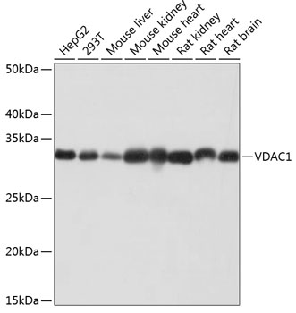
Western blot analysis of various lysates using VDAC1 Rabbit mAb (A19707) at 1:6000 dilution incubated overnight at 4°C. Secondary antibody: HRP-conjugated Goat anti-Rabbit IgG (H+L) (AS014) at 1:10000 dilution. Lysates/proteins: 25 µg per lane. Blocking buffer: 3% nonfat dry milk in TBST. Detection: ECL Basic Kit (RM00020). Exposure time: 10s. |
|
|
Confocal imaging of L-929 cells using VDAC1 Rabbit mAb (A19707, dilution 1:200) followed by a further incubation with Cy3 Goat Anti-Rabbit IgG (H+L) (AS007, dilution 1:500) (Red). The cells were counterstained with alpha-Tubulin Mouse mAb (AC012, dilution 1:400) followed by incubation with ABflo 488-conjugated Goat Anti-Mouse IgG (H+L) Ab (AS076, dilution 1:500) (Green). DAPI was used for nuclear staining (Blue). Objective: 100x. |
|
|
Confocal imaging of NIH/3T3 cells using VDAC1 Rabbit mAb (A19707, dilution 1:200) followed by a further incubation with Cy3 Goat Anti-Rabbit IgG (H+L) (AS007, dilution 1:500) (Red). The cells were counterstained with alpha-Tubulin Mouse mAb (AC012, dilution 1:400) followed by incubation with ABflo 488-conjugated Goat Anti-Mouse IgG (H+L) Ab (AS076, dilution 1:500) (Green). DAPI was used for nuclear staining (Blue). Objective: 100x. |
|
|
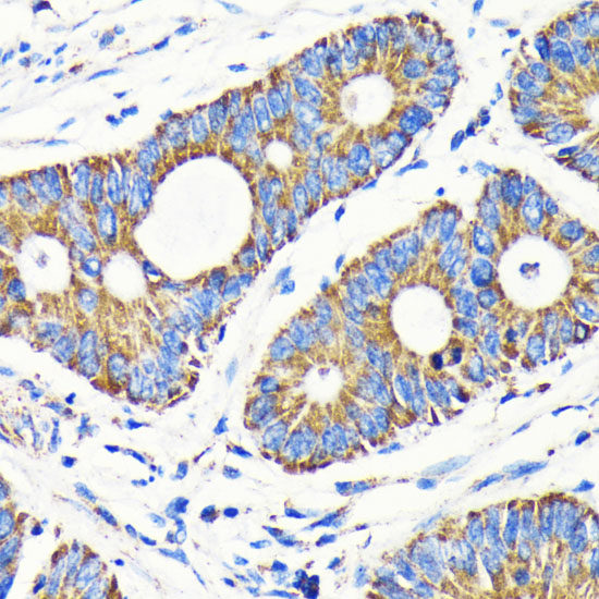
Immunohistochemistry analysis of paraffin-embedded Human colon carcinoma tissue using VDAC1 Rabbit mAb (A19707) at a dilution of 1:2000 (40x lens). High pressure antigen retrieval performed with 0.01M Tris-EDTA Buffer (pH 9.0) prior to IHC staining. |
|
|
Immunohistochemistry analysis of paraffin-embedded Human pancreas tissue using VDAC1 Rabbit mAb (A19707) at a dilution of 1:2000 (40x lens). High pressure antigen retrieval performed with 0.01M Tris-EDTA Buffer (pH 9.0) prior to IHC staining. |
|
|
Immunohistochemistry analysis of paraffin-embedded Mouse testis tissue using VDAC1 Rabbit mAb (A19707) at a dilution of 1:2000 (40x lens). High pressure antigen retrieval performed with 0.01M Tris-EDTA Buffer (pH 9.0) prior to IHC staining. |
|
|
Immunohistochemistry analysis of paraffin-embedded Rat liver tissue using VDAC1 Rabbit mAb (A19707) at a dilution of 1:2000 (40x lens). High pressure antigen retrieval performed with 0.01M Tris-EDTA Buffer (pH 9.0) prior to IHC staining. |
Produktgarantie und fachkundiger Support
