
HINT1 Rabbit mAb, Unconjugated, Monoclonal
Artikelnummer:
ABB-A20894
- Bilder (9)
| Artikelname: | HINT1 Rabbit mAb, Unconjugated, Monoclonal |
| Artikelnummer: | ABB-A20894 |
| Hersteller Artikelnummer: | A20894 |
| Alternativnummer: | ABB-A20894-20UL,ABB-A20894-100UL |
| Hersteller: | ABclonal |
| Wirt: | Rabbit |
| Kategorie: | Antikörper |
| Applikation: | ELISA, IF, IHC-P, WB |
| Spezies Reaktivität: | Human |
| Immunogen: | Synthetic peptide. This information is considered to be commercially sensitive. |
| Konjugation: | Unconjugated |
| Alternative Synonym: | HINT, NMAN, PKCI-1, PRKCNH1, HINT1 |
| This gene encodes a protein that hydrolyzes purine nucleotide phosphoramidates substrates, including AMP-morpholidate, AMP-N-alanine methyl ester, AMP-alpha-acetyl lysine methyl ester, and AMP-NH2. The encoded protein interacts with these substrates via a histidine triad motif. This gene is considered a tumor suppressor gene. In addition, mutations in this gene can cause autosomal recessive neuromyotonia and axonal neuropathy. There are several related pseudogenes on chromosome 7. Several transcript variants have been observed. |
| Application Verdünnung: | WB,1:500 - 1:1000|IHC-P,1:50 - 1:200|IF/ICC,1:50 - 1:200|ELISA,Recommended starting concentration is 1 µg/mL. Please optimize the concentration based on your specific assay requirements. |
| Anwendungsbeschreibung: | Cross-Reactivity: Human,Mouse,Rat. ResearchArea: Epigenetics Nuclear Signaling,Cancer,Tumor suppressors. Shipping: Ice Bag |
|
|
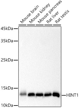
Western blot analysis of various lysates using HINT1 Rabbit mAb (A20894) at 1:1000 dilution. Secondary antibody: HRP-conjugated Goat anti-Rabbit IgG (H+L) (AS014) at 1:10000 dilution. Lysates/proteins: 25µg per lane. Blocking buffer: 3% nonfat dry milk in TBST. Detection: ECL Basic Kit (RM00020). Exposure time: 3s. |
|
|
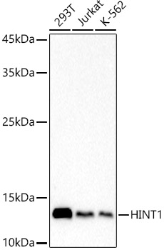
Western blot analysis of various lysates using HINT1 Rabbit mAb (A20894) at 1:1000 dilution. Secondary antibody: HRP-conjugated Goat anti-Rabbit IgG (H+L) (AS014) at 1:10000 dilution. Lysates/proteins: 25µg per lane. Blocking buffer: 3% nonfat dry milk in TBST. Detection: ECL Basic Kit (RM00020). Exposure time: 10s. |
|
|
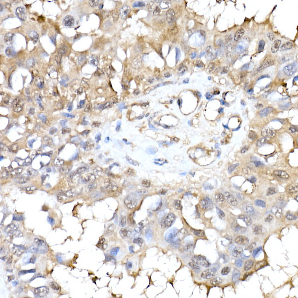
Immunohistochemistry analysis of paraffin-embedded Human breast cancer using HINT1 Rabbit mAb (A20894) at dilution of 1:50 (40x lens). High pressure antigen retrieval performed with 0.01M Citrate buffer (pH 6.0) prior to IHC staining. |
|
|
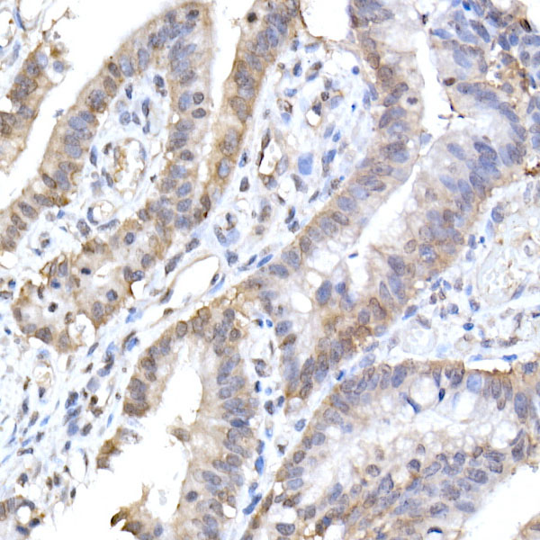
Immunohistochemistry analysis of paraffin-embedded Human colon carcinoma using HINT1 Rabbit mAb (A20894) at dilution of 1:50 (40x lens). High pressure antigen retrieval performed with 0.01M Citrate buffer (pH 6.0) prior to IHC staining. |
|
|
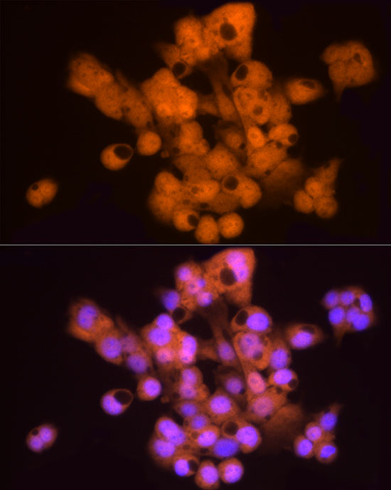
Immunofluorescence analysis of HepG2 cells using HINT1 Rabbit mAb (A20894) at dilution of 1:50 (40x lens). Secondary antibody: Cy3-conjugated Goat anti-Rabbit IgG (H+L) (AS007) at 1:500 dilution. Blue: DAPI for nuclear staining. |
|
|
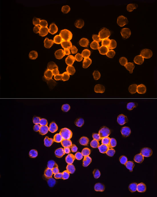
Immunofluorescence analysis of Jurkat cells using HINT1 Rabbit mAb (A20894) at dilution of 1:50 (40x lens). Secondary antibody: Cy3-conjugated Goat anti-Rabbit IgG (H+L) (AS007) at 1:500 dilution. Blue: DAPI for nuclear staining. |
|
|
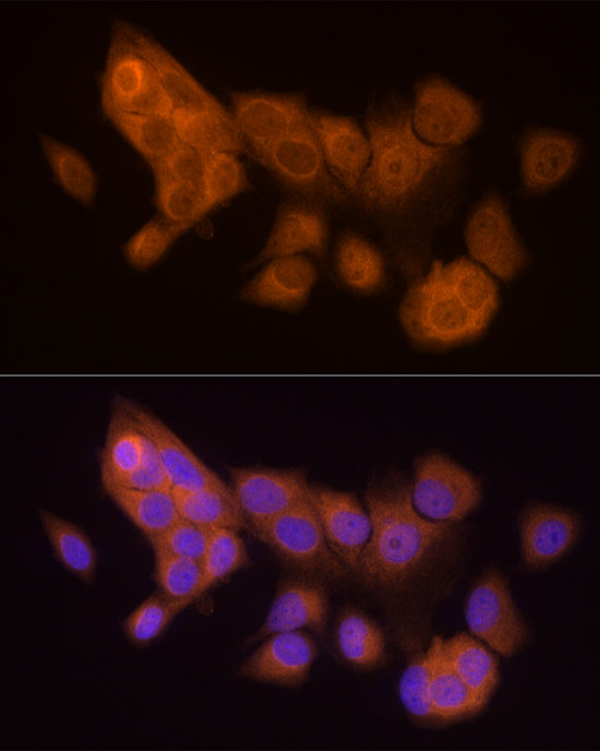
Immunofluorescence analysis of MCF7 cells using HINT1 Rabbit mAb (A20894) at dilution of 1:50 (40x lens). Secondary antibody: Cy3-conjugated Goat anti-Rabbit IgG (H+L) (AS007) at 1:500 dilution. Blue: DAPI for nuclear staining. |
|
|
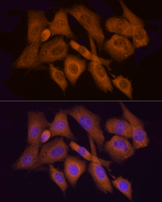
Immunofluorescence analysis of NIH/3T3 cells using HINT1 Rabbit mAb (A20894) at dilution of 1:50 (40x lens). Secondary antibody: Cy3-conjugated Goat anti-Rabbit IgG (H+L) (AS007) at 1:500 dilution. Blue: DAPI for nuclear staining. |
|
|
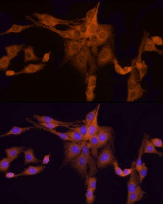
Immunofluorescence analysis of PC-12 cells using HINT1 Rabbit mAb (A20894) at dilution of 1:50 (40x lens). Secondary antibody: Cy3-conjugated Goat anti-Rabbit IgG (H+L) (AS007) at 1:500 dilution. Blue: DAPI for nuclear staining. |
Produktgarantie und fachkundiger Support
