
[KO Validated] Non-phospho (Active)beta-Catenin -S33/S37/T41 Rabbit mAb, Unconjugated, Monoclonal
Artikelnummer:
ABB-A22180
- Bilder (9)

| Artikelname: | [KO Validated] Non-phospho (Active)beta-Catenin -S33/S37/T41 Rabbit mAb, Unconjugated, Monoclonal |
| Artikelnummer: | ABB-A22180 |
| Hersteller Artikelnummer: | A22180 |
| Alternativnummer: | ABB-A22180-100UL,ABB-A22180-20UL,ABB-A22180-500UL,ABB-A22180-1000UL |
| Hersteller: | ABclonal |
| Wirt: | Rabbit |
| Kategorie: | Antikörper |
| Applikation: | ELISA, IHC-P, WB |
| Spezies Reaktivität: | Human |
| Immunogen: | Synthetic peptide. This information is considered to be commercially sensitive. |
| Konjugation: | Unconjugated |
| Alternative Synonym: | EVR7, CTNNB, MRD19, NEDSDV, armadillo, Non-phospho (Active)beta-Catenin -S33/S37/T41 |
| The protein encoded by this gene is part of a complex of proteins that constitute adherens junctions (AJs). AJs are necessary for the creation and maintenance of epithelial cell layers by regulating cell growth and adhesion between cells. The encoded protein also anchors the actin cytoskeleton and may be responsible for transmitting the contact inhibition signal that causes cells to stop dividing once the epithelial sheet is complete. Finally, this protein binds to the product of the APC gene, which is mutated in adenomatous polyposis of the colon. Mutations in this gene are a cause of colorectal cancer (CRC), pilomatrixoma (PTR), medulloblastoma (MDB), and ovarian cancer. Alternative splicing results in multiple transcript variants. |
| Klonalität: | Monoclonal |
| Klon-Bezeichnung: | [ARC53641] |
| Molekulargewicht: | 85kDa |
| NCBI: | 1499 |
| UniProt: | P35222 |
| Reinheit: | Affinity purification |
| Sequenz: | MATQADLMELDMAMEPDRKAAVSHWQQQSYLDSGIHSGATTTAPSLSGKGNPEEEDVDTSQVLYEWEQGFSQSFTQEQVADIDGQYAMTRAQRVRAAMFP |
| Target-Kategorie: | CTNNB1 |
| Antibody Type: | Primary Antibody |
| Application Verdünnung: | WB,1:2000 - 1:10000|IHC-P,1:1000 - 1:4000|ELISA,Recommended starting concentration is 1 µg/mL. Please optimize the concentration based on your specific assay requirements. |
| Anwendungsbeschreibung: | Cross-Reactivity: Human,Mouse,Rat. ResearchArea: Epigenetics Nuclear Signaling,Transcription Factors,Protein phosphorylation,Cancer,Invasion and Metastasis,Signal Transduction,ErbB-HER Signaling Pathway,Cell Biology Developmental Biology,Apoptosis,Cell Adhesion,Cadherins,Tight Junctions,Cytoskeleton,Microfilaments,Wnt -Catenin Signaling Pathway,ESC Pluripotency and Differentiation,Immunology Inflammation,Neuroscience,Stem Cells,Cardiovascular,Angiogenesis. Shipping: Ice Bag |
|
|
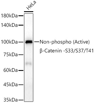
Western blot analysis of lysates from wild type (WT) and Non-phospho (Active)beta-Catenin -S33/S37/T41 knockout (KO) 293T cells using [KO Validated] Non-phospho (Active)beta-Catenin -S33/S37/T41 Rabbit mAb (A22180) at 1:16000 dilution incubated overnight at 4°C. Secondary antibody: HRP-conjugated Goat anti-Rabbit IgG (H+L) (AS014) at 1:10000 dilution. Lysates/proteins: 25 µg per lane. Blocking buffer: 3% nonfat dry milk in TBST. Detection: ECL Basic Kit (RM00020). Exposure time: 90s. |
|
|
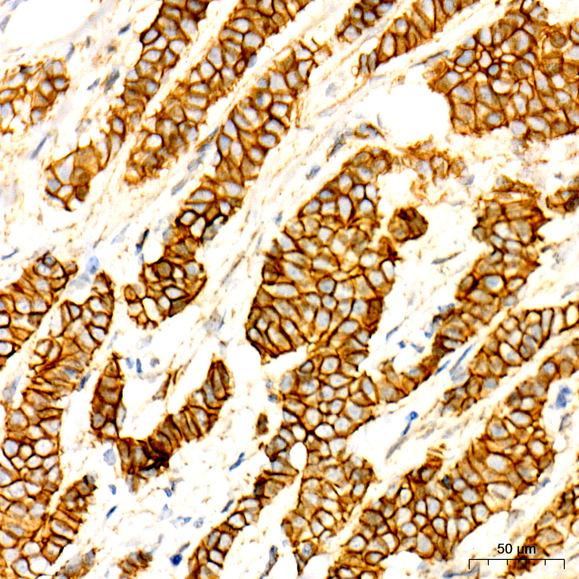
Immunohistochemistry analysis of paraffin-embedded Human placenta tissue using [KO Validated] Non-phospho (Active)beta-Catenin -S33/S37/T41 Rabbit mAb (A22180) at a dilution of 1:1500 (40x lens). High pressure antigen retrieval performed with 0.01M Tris-EDTA Buffer (pH 9.0) prior to IHC staining. |
|
|
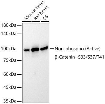
Western blot analysis of various lysates using [KO Validated] Non-phospho (Active)beta-Catenin -S33/S37/T41 Rabbit mAb (A22180) at 1:11000 dilution incubated overnight at 4°C. Secondary antibody: HRP-conjugated Goat anti-Rabbit IgG (H+L) (AS014) at 1:10000 dilution. Lysates/proteins: 25 µg per lane. Blocking buffer: 3% nonfat dry milk in TBST. Detection: ECL Basic Kit (RM00020). Exposure time: 20s. |
|
|
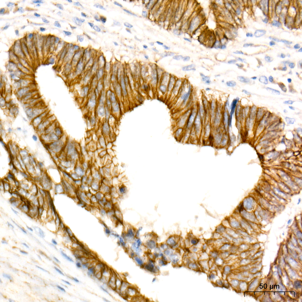
Immunohistochemistry analysis of paraffin-embedded Human pancreas tissue using [KO Validated] Non-phospho (Active)beta-Catenin -S33/S37/T41 Rabbit mAb (A22180) at a dilution of 1:1500 (40x lens). High pressure antigen retrieval performed with 0.01M Tris-EDTA Buffer (pH 9.0) prior to IHC staining. |
|
|
Immunohistochemistry analysis of paraffin-embedded Human stomach tissue using [KO Validated] Non-phospho (Active)beta-Catenin -S33/S37/T41 Rabbit mAb (A22180) at a dilution of 1:1500 (40x lens). High pressure antigen retrieval performed with 0.01M Tris-EDTA Buffer (pH 9.0) prior to IHC staining. |
|
|
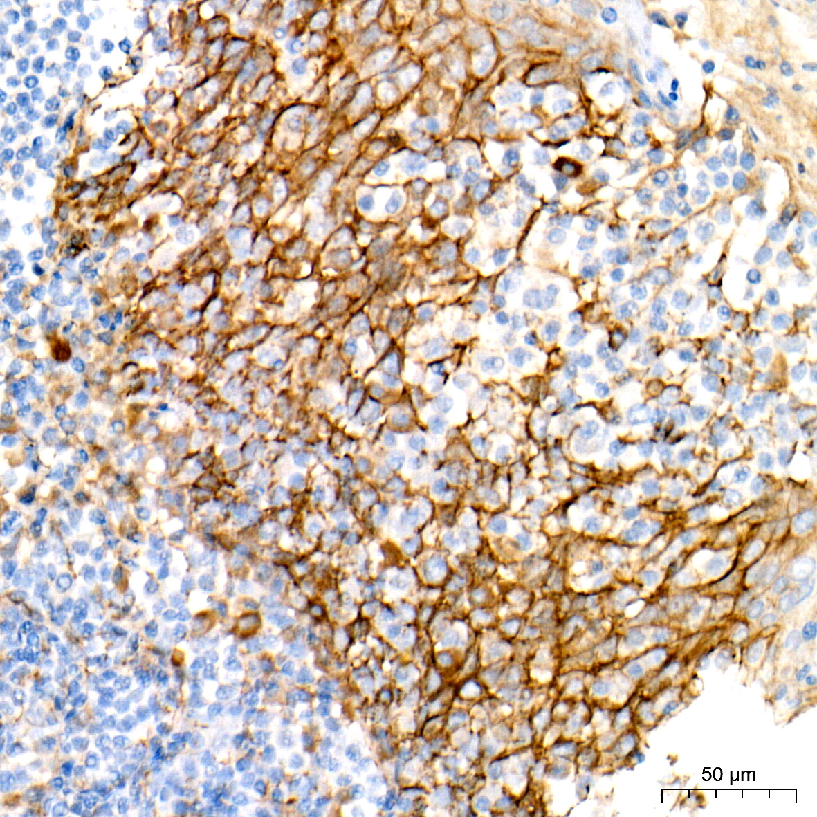
Immunohistochemistry analysis of paraffin-embedded Human thyroid cancer tissue using [KO Validated] Non-phospho (Active)beta-Catenin -S33/S37/T41 Rabbit mAb (A22180) at a dilution of 1:1500 (40x lens). High pressure antigen retrieval performed with 0.01M Tris-EDTA Buffer (pH 9.0) prior to IHC staining. |
|
|
Immunohistochemistry analysis of paraffin-embedded Mouse intestin tissue using [KO Validated] Non-phospho (Active)beta-Catenin -S33/S37/T41 Rabbit mAb (A22180) at a dilution of 1:1500 (40x lens). High pressure antigen retrieval performed with 0.01M Tris-EDTA Buffer (pH 9.0) prior to IHC staining. |
|
|
Immunohistochemistry analysis of paraffin-embedded Mouse pancreas tissue using [KO Validated] Non-phospho (Active)beta-Catenin -S33/S37/T41 Rabbit mAb (A22180) at a dilution of 1:1500 (40x lens). High pressure antigen retrieval performed with 0.01M Tris-EDTA Buffer (pH 9.0) prior to IHC staining. |
|
|
Immunohistochemistry analysis of paraffin-embedded Rat colon tissue using [KO Validated] Non-phospho (Active)beta-Catenin -S33/S37/T41 Rabbit mAb (A22180) at a dilution of 1:1500 (40x lens). High pressure antigen retrieval performed with 0.01M Tris-EDTA Buffer (pH 9.0) prior to IHC staining. |
Produktgarantie und fachkundiger Support
