
MonoMethyl-Histone H4-K20 Rabbit mAb, Unconjugated
Artikelnummer:
ABB-A22572
- Bilder (9)

| Artikelname: | MonoMethyl-Histone H4-K20 Rabbit mAb, Unconjugated |
| Artikelnummer: | ABB-A22572 |
| Hersteller Artikelnummer: | A22572 |
| Alternativnummer: | ABB-A22572-100UL,ABB-A22572-20UL,ABB-A22572-1000UL,ABB-A22572-500UL |
| Hersteller: | ABclonal |
| Wirt: | Rabbit |
| Kategorie: | Antikörper |
| Applikation: | DOT, ELISA, IHC-P, WB |
| Spezies Reaktivität: | Human |
| Immunogen: | Synthetic peptide. This information is considered to be commercially sensitive. |
| Konjugation: | Unconjugated |
| Alternative Synonym: | H4, H4/n, H4C1, H4C2, H4C3, H4C4, H4C5, H4C6, H4C8, H4C9, H4F2, H4FN, FO108, H4-16, H4C11, H4C12, H4C13, H4C15, H4C16, HIST2H4, HIST2H4A, MonoMethyl-Histone H4-K20 |
| Histones are basic nuclear proteins that are responsible for the nucleosome structure of the chromosomal fiber in eukaryotes. This structure consists of approximately 146 bp of DNA wrapped around a nucleosome, an octamer composed of pairs of each of the four core histones (H2A, H2B, H3, and H4). The chromatin fiber is further compacted through the interaction of a linker histone, H1, with the DNA between the nucleosomes to form higher order chromatin structures. This gene is intronless and encodes a replication-dependent histone that is a member of the histone H4 family. Transcripts from this gene lack polyA tails, instead, they contain a palindromic termination element. This gene is found in a histone cluster on chromosome 1. This gene is one of four histone genes in the cluster that are duplicated, this record represents the centromeric copy. |
| Klonalität: | Monoclonal |
| Klon-Bezeichnung: | [ARC54049] |
| Molekulargewicht: | 11kDa |
| NCBI: | 8359 |
| UniProt: | P62805 |
| Reinheit: | Affinity purification |
| Sequenz: | MSGRGKGGKGLGKGGAKRHRKVLRDNIQGITKPAIRRLARRGGVKRISGLIYEETRGVLKVFLENVIRDAVTYTEHAKRKTVTAMDVVYALKRQGRTLYG |
| Target-Kategorie: | Histone H4 |
| Antibody Type: | Primary Antibody |
| Application Verdünnung: | WB,1:500 - 1:1000|DB,1:500 - 1:1000|IHC-P,1:100 - 1:500|ELISA,Recommended starting concentration is 1 µg/mL. Please optimize the concentration based on your specific assay requirements. |
| Anwendungsbeschreibung: | Cross-Reactivity: Human,Mouse,Rat,Other (Wide Range Predicted). ResearchArea: Epigenetics Nuclear Signaling. Shipping: Ice Bag |
|
|
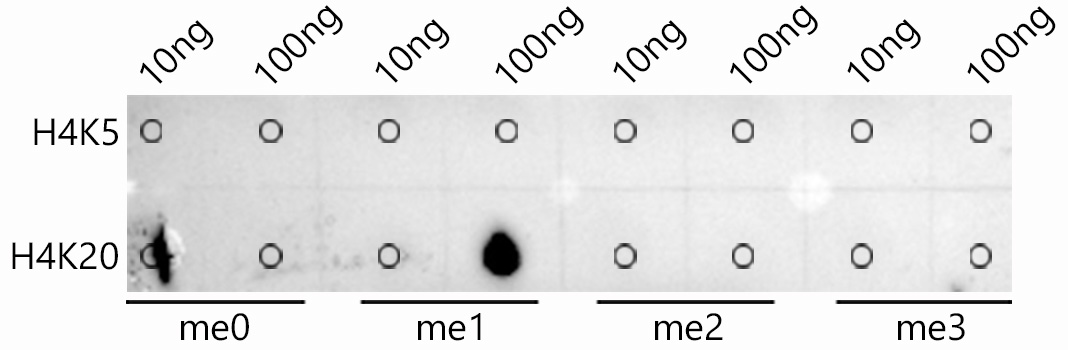
Dot-blot analysis of all sorts of peptides using MonoMethyl-Histone H4-K20 antibody (A22572) at 1:1000 dilution. |
|
|
Immunohistochemistry analysis of paraffin-embeddedRat testis tissue usingMonoMethyl-Histone H4-K20 Rabbit mAb(A22572) at a dilution of 1:400 (40x lens).High pressure antigen retrieval was performed with 0.01 M citrate buffer (pH 6.0) prior to IHC staining. |
|
|
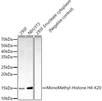
Western blot analysis of various lysates, using MonoMethyl-Histone H4-K20 Rabbit mAb (A22572) at 1:900 dilution. Secondary antibody: HRP-conjugated Goat anti-Rabbit IgG (H+L) (AS014) at 1:10000 dilution. Lysates/proteins: 25µg per lane. Blocking buffer: 3% nonfat dry milk in TBST. Detection: ECL Enhanced Kit (RM00021). Exposure time: 180s. |
|
|
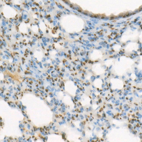
Immunohistochemistry analysis of paraffin-embeddedRat spleen tissue usingMonoMethyl-Histone H4-K20 Rabbit mAb(A22572) at a dilution of 1:400 (40x lens).High pressure antigen retrieval was performed with 0.01 M citrate buffer (pH 6.0) prior to IHC staining. |
|
|
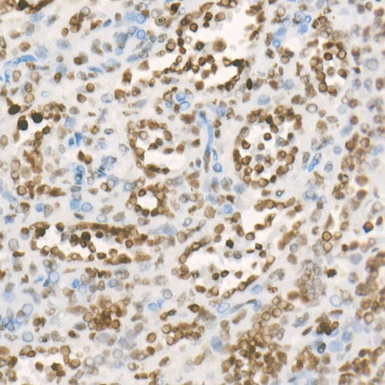
Immunohistochemistry analysis of paraffin-embeddedMouse testis tissue usingMonoMethyl-Histone H4-K20 Rabbit mAb(A22572) at a dilution of 1:400 (40x lens).High pressure antigen retrieval was performed with 0.01 M citrate buffer (pH 6.0) prior to IHC staining. |
|
|
Immunohistochemistry analysis of paraffin-embeddedMouse brain tissue usingMonoMethyl-Histone H4-K20 Rabbit mAb(A22572) at a dilution of 1:400 (40x lens).High pressure antigen retrieval was performed with 0.01 M citrate buffer (pH 6.0) prior to IHC staining. |
|
|
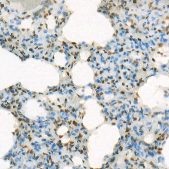
Immunohistochemistry analysis of paraffin-embeddedRat lung tissue usingMonoMethyl-Histone H4-K20 Rabbit mAb(A22572) at a dilution of 1:400 (40x lens).High pressure antigen retrieval was performed with 0.01 M citrate buffer (pH 6.0) prior to IHC staining. |
|
|
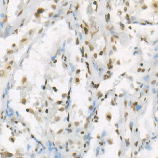
Immunohistochemistry analysis of paraffin-embeddedHuman breast cancer tissue usingMonoMethyl-Histone H4-K20 Rabbit mAb(A22572) at a dilution of 1:400 (40x lens).High pressure antigen retrieval was performed with 0.01 M citrate buffer (pH 6.0) prior to IHC staining. |
|
|
Immunohistochemistry analysis of paraffin-embeddedMouse kidney tissue usingMonoMethyl-Histone H4-K20 Rabbit mAb(A22572) at a dilution of 1:400 (40x lens).High pressure antigen retrieval was performed with 0.01 M citrate buffer (pH 6.0) prior to IHC staining. |
Produktgarantie und fachkundiger Support
