
GSK-3alpha/beta Rabbit mAb, Unconjugated, Monoclonal
Artikelnummer:
ABB-A22666
- Bilder (9)

| Artikelname: | GSK-3alpha/beta Rabbit mAb, Unconjugated, Monoclonal |
| Artikelnummer: | ABB-A22666 |
| Hersteller Artikelnummer: | A22666 |
| Alternativnummer: | ABB-A22666-20UL,ABB-A22666-100UL |
| Hersteller: | ABclonal |
| Wirt: | Rabbit |
| Kategorie: | Antikörper |
| Applikation: | ELISA, IF, IHC-P, WB |
| Spezies Reaktivität: | Human |
| Immunogen: | Recombinant protein (or fragment).This information is considered to be commercially sensitive. |
| Konjugation: | Unconjugated |
| Alternative Synonym: | GSK3B, gsk-3beta, GSK-3alpha/beta |
| The protein encoded by this gene is a serine-threonine kinase, belonging to the glycogen synthase kinase subfamily. It is involved in energy metabolism, neuronal cell development, and body pattern formation. Polymorphisms in this gene have been implicated in modifying risk of Parkinson disease, and studies in mice show that overexpression of this gene may be relevant to the pathogenesis of Alzheimer disease. Alternatively spliced transcript variants encoding different isoforms have been found for this gene. |
| Application Verdünnung: | WB,1:10000 - 1:60000|IF/ICC,1:50 - 1:200|IF-P,1:50 - 1:200|IHC-P,1:500- 1:1000|ELISA,Recommended starting concentration is 1 µg/mL. Please optimize the concentration based on your specific assay requirements. |
| Anwendungsbeschreibung: | Cross-Reactivity: Human,Mouse,Rat. ResearchArea: Epigenetics & Nuclear Signaling,Translation Control,Regulation of eIF4 and p70 S6 Kinase,Regulation of eIF2,Cancer,Signal Transduction,Kinase,Serine/threonine kinases,PI3K-Akt Signaling Pathway,mTOR Signaling Pathway,ErbB-HER Signaling Pathway,MAPK-Erk Signaling Pathway,Cell Biology & Developmental Biology,Apoptosis,Inhibition of Apoptosis,Cell Cycle,Centrosome,G1/S Checkpoint,Cell Adhesion,Microtubules,Hedgehog Signaling Pathway,Wnt/beta-Catenin Signaling Pathway,ESC Pluripotency and Differentiation,Endocrine & Metabolism,Carbohydrate metabolism,Insulin Receptor Signaling Pathway,Endocrine and metabolic diseases,Diabetes,Immunology & Inflammation,B Cell Receptor Signaling Pathway,NF-kB Signaling Pathway,Neuroscience,Neurodegenerative Diseases,Amyloid Plaque and Neurofibrillary Tangle Formation in Alzheimers Disease,Neurodegenerative Diseases Markers,Other Neurological disorders,Stem Cells,Cardiovascular,Heart,Hypertrophy,Akt downstream targets,Protein phosphorylation. Shipping: Ice Bag |
|
|
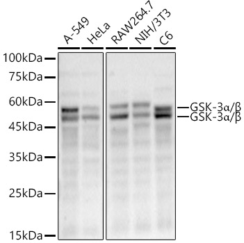
Western blot analysis of various lysates, using GSK-3alpha/beta Rabbit mAb (A22666) at 1:50000 dilution. Secondary antibody: HRP-conjugated Goat anti-Rabbit IgG (H+L) (AS014) at 1:10000 dilution. Lysates/proteins: 25µg per lane. Blocking buffer: 3% nonfat dry milk in TBST. Detection: ECL Enhanced Kit (RM00021). Exposure time: 90s. |
|
|
Immunohistochemistry analysis of paraffin-embeddedMouse colon tissue usingGSK-3alpha/beta Rabbit mAb(A22666) at a dilution of 1:800 (40x lens).High pressure antigen retrieval was performed with 0.01 M citrate buffer (pH 6.0) prior to IHC staining. |
|
|
Immunohistochemistry analysis of paraffin-embeddedHuman liver tissue usingGSK-3alpha/beta Rabbit mAb(A22666) at a dilution of 1:800 (40x lens).High pressure antigen retrieval was performed with 0.01 M citrate buffer (pH 6.0) prior to IHC staining. |
|
|
Immunohistochemistry analysis of paraffin-embeddedMouse kidney tissue usingGSK-3alpha/beta Rabbit mAb(A22666) at a dilution of 1:800 (40x lens).High pressure antigen retrieval was performed with 0.01 M citrate buffer (pH 6.0) prior to IHC staining. |
|
|
Immunohistochemistry analysis of paraffin-embeddedRat colon tissue usingGSK-3alpha/beta Rabbit mAb(A22666) at a dilution of 1:800 (40x lens).High pressure antigen retrieval was performed with 0.01 M citrate buffer (pH 6.0) prior to IHC staining. |
|
|
Immunohistochemistry analysis of paraffin-embeddedMouse testis tissue usingGSK-3alpha/beta Rabbit mAb(A22666) at a dilution of 1:800 (40x lens).High pressure antigen retrieval was performed with 0.01 M citrate buffer (pH 6.0) prior to IHC staining. |
|
|
Confocal imaging of HeLa cells using GSK-3alpha/beta Rabbit mAb (A22666, dilution 1:200) followed by a further incubation with Cy3 Goat Anti-Rabbit IgG (H+L) (AS007, dilution 1:500) (Red). The cells were counterstained with alpha-Tubulin Mouse mAb (AC012, dilution 1:400) followed by incubation with ABflo 488-conjugated Goat Anti-Mouse IgG (H+L) Ab (AS076, dilution 1:500) (Green). DAPI was used for nuclear staining (Blue). Objective: 100x. |
|
|
Confocal imaging of paraffin-embedded Mouse brain tissue using GSK-3alpha/beta Rabbit mAb (A22666, dilution 1:200) followed by a further incubation with Cy3 Goat Anti-Rabbit IgG (H+L) (AS007, dilution 1:500) (Red). DAPI was used for nuclear staining (Blue). Microwave antigen retrieval performed with 0.01M Citrate Buffer (pH 6.0) prior to IF staining. Objective: 40x. |
|
|
Confocal imaging of paraffin-embedded Rat brain tissue using GSK-3alpha/beta Rabbit mAb (A22666, dilution 1:200) followed by a further incubation with Cy3 Goat Anti-Rabbit IgG (H+L) (AS007, dilution 1:500) (Red). DAPI was used for nuclear staining (Blue). Microwave antigen retrieval performed with 0.01M Citrate Buffer (pH 6.0) prior to IF staining. Objective: 40x. |
Produktgarantie und fachkundiger Support
