
TFAM Rabbit mAb, Unconjugated, Monoclonal
Artikelnummer:
ABB-A23467
- Bilder (9)

| Artikelname: | TFAM Rabbit mAb, Unconjugated, Monoclonal |
| Artikelnummer: | ABB-A23467 |
| Hersteller Artikelnummer: | A23467 |
| Alternativnummer: | ABB-A23467-20UL,ABB-A23467-500UL,ABB-A23467-100UL,ABB-A23467-1000UL |
| Hersteller: | ABclonal |
| Wirt: | Rabbit |
| Kategorie: | Antikörper |
| Applikation: | ChIP, ELISA, IF, IHC-P, IP, WB |
| Spezies Reaktivität: | Human |
| Immunogen: | Recombinant protein (or fragment).This information is considered to be commercially sensitive. |
| Konjugation: | Unconjugated |
| Alternative Synonym: | TCF6, MTTF1, MTTFA, TCF6L1, TCF6L2, TCF6L3, MTDPS15, TFAM |
| This gene encodes a key mitochondrial transcription factor containing two high mobility group motifs. The encoded protein also functions in mitochondrial DNA replication and repair. Sequence polymorphisms in this gene are associated with Alzheimers and Parkinsons diseases. There are pseudogenes for this gene on chromosomes 6, 7, and 11. Alternative splicing results in multiple transcript variants. |
| Application Verdünnung: | WB,1:2000 - 1:8000|IHC-P,1:50 - 1:200|IF/ICC,1:50 - 1:200|IP,0.5µg-4µg antibody for 200µg-400µg extracts of whole cells|ELISA,Recommended starting concentration is 1 µg/mL. Please optimize the concentration based on your specific assay requirements.|ChIP, |
| Anwendungsbeschreibung: | Cross-Reactivity: Human. ResearchArea: Epigenetics Nuclear Signaling,Transcription Factors,RNA Binding,Cancer,Signal Transduction,Cell Biology Developmental Biology,Cell Adhesion,Wnt -Catenin Signaling Pathway,Endocrine Metabolism,Mitochondrial metabolism,Mitochondrial markers,Nucleotide metabolism,Neuroscience,Neurodegenerative Diseases,Cardiovascular,Heart,Cardiogenesis. Shipping: Ice Bag |
|
|
Western blot analysis of lysates from 293F cells using TFAM Rabbit mAb (A23467) at 1:4000 dilutionincubated overnight at 4°C. Mitochondrial extracts derived from 293F cells. Secondary antibody: HRP-conjugated Goat anti-Rabbit IgG (H+L) (AS014) at 1:10000 dilution. Lysates/proteins: 25 µg per lane. Blocking buffer: 3% nonfat dry milk in TBST. Detection: ECL Basic Kit (RM00020). Exposure time: 1 s. |
|
|
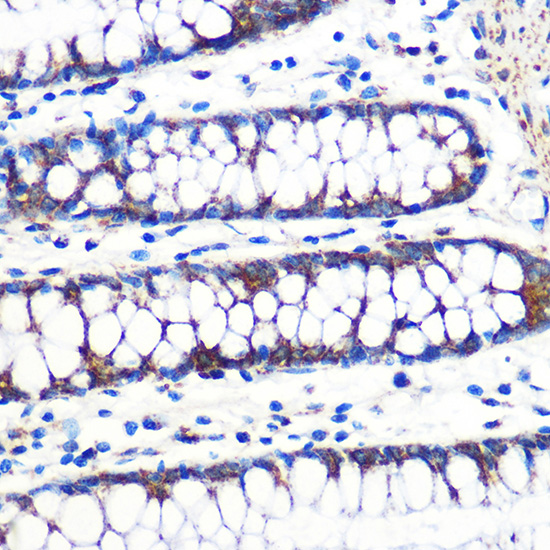
Immunohistochemistry analysis of paraffin-embeddedHuman brain tissue usingTFAM Rabbit mAb(A23467) at a dilution of 1:200 (40x lens).High pressure antigen retrieval was performed with 0.01 M citrate buffer (pH 6.0) prior to IHC staining. |
|
|
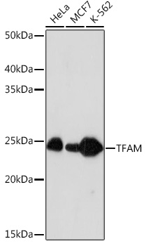
Western blot analysis of various lysates using TFAM Rabbit mAb (A23467) at 1:4000 dilution incubated overnight at 4°C. Secondary antibody: HRP-conjugated Goat anti-Rabbit IgG (H+L) (AS014) at 1:10000 dilution. Lysates/proteins: 25 µg per lane. Blocking buffer: 3% nonfat dry milk in TBST. Detection: ECL Basic Kit (RM00020). Exposure time: 30 s. |
|
|
Immunohistochemistry analysis of paraffin-embeddedHuman kidney tissue usingTFAM Rabbit mAb(A23467) at a dilution of 1:200 (40x lens).High pressure antigen retrieval was performed with 0.01 M citrate buffer (pH 6.0) prior to IHC staining. |
|
|
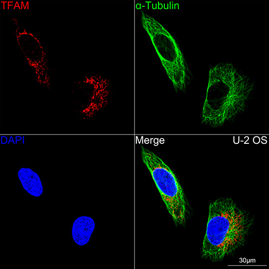
Confocal imaging of U-2 OS cells using TFAM Rabbit mAb (A23467, dilution 1:200) followed by a further incubation with Cy3 Goat Anti-Rabbit IgG (H+L) (AS007, dilution 1:500) (Red). The cells were counterstained with alpha-Tubulin Mouse mAb (AC012, dilution 1:400) followed by incubation with ABflo 488-conjugated Goat Anti-Mouse IgG (H+L) Ab (AS076, dilution 1:500) (Green). DAPI was used for nuclear staining (Blue). Objective: 100x. |
|
|
Confocal imaging of HeLa cells using TFAM Rabbit mAb (A23467, dilution 1:200) followed by a further incubation with Cy3 Goat Anti-Rabbit IgG (H+L) (AS007, dilution 1:500) (Red). The cells were counterstained with alpha-Tubulin Mouse mAb (AC012, dilution 1:400) followed by incubation with ABflo 488-conjugated Goat Anti-Mouse IgG (H+L) Ab (AS076, dilution 1:500) (Green). DAPI was used for nuclear staining (Blue). Objective: 100x. |
|
|
Confocal imaging of Hep G2 cells using TFAM Rabbit mAb (A23467, dilution 1:200) followed by a further incubation with Cy3 Goat Anti-Rabbit IgG (H+L) (AS007, dilution 1:500) (Red). The cells were counterstained with alpha-Tubulin Mouse mAb (AC012, dilution 1:400) followed by incubation with ABflo 488-conjugated Goat Anti-Mouse IgG (H+L) Ab (AS076, dilution 1:500) (Green). DAPI was used for nuclear staining (Blue). Objective: 100x. |
|
|
Confocal imaging of MCF7 cells using TFAM Rabbit mAb (A23467, dilution 1:200) followed by a further incubation with Cy3 Goat Anti-Rabbit IgG (H+L) (AS007, dilution 1:500) (Red). The cells were counterstained with alpha-Tubulin Mouse mAb (AC012, dilution 1:400) followed by incubation with ABflo 488-conjugated Goat Anti-Mouse IgG (H+L) Ab (AS076, dilution 1:500) (Green). DAPI was used for nuclear staining (Blue). Objective: 100x. |
|
|
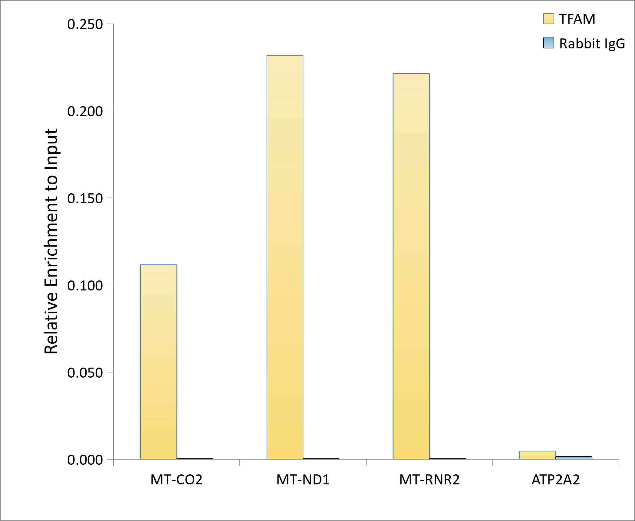
Chromatin immunoprecipitation analysis of extracts of K-562 cells, using TFAM Rabbit mAb (A23467) and rabbit IgG.The amount of immunoprecipitated DNA was checked by quantitative PCR. Histogram was constructed by the ratios of the immunoprecipitated DNA to the input. |
Produktgarantie und fachkundiger Support
