
Integrin-beta1/CD29 Rabbit mAb, Unconjugated, Monoclonal
Artikelnummer:
ABB-A23497
- Bilder (9)

| Artikelname: | Integrin-beta1/CD29 Rabbit mAb, Unconjugated, Monoclonal |
| Artikelnummer: | ABB-A23497 |
| Hersteller Artikelnummer: | A23497 |
| Alternativnummer: | ABB-A23497-100UL,ABB-A23497-1000UL,ABB-A23497-500UL,ABB-A23497-20UL |
| Hersteller: | ABclonal |
| Wirt: | Rabbit |
| Kategorie: | Antikörper |
| Applikation: | ELISA, FC, IF, IHC-P, WB |
| Spezies Reaktivität: | Human |
| Immunogen: | Recombinant protein (or fragment).This information is considered to be commercially sensitive. |
| Konjugation: | Unconjugated |
| Alternative Synonym: | CD29, FNRB, MDF2, VLAB, GPIIA, MSK12, VLA-BETA, Integrin-beta1/CD29 |
| Integrins are heterodimeric proteins made up of alpha and beta subunits. At least 18 alpha and 8 beta subunits have been described in mammals. Integrin family members are membrane receptors involved in cell adhesion and recognition in a variety of processes including embryogenesis, hemostasis, tissue repair, immune response and metastatic diffusion of tumor cells. This gene encodes a beta subunit. Multiple alternatively spliced transcript variants which encode different protein isoforms have been found for this gene. |
| Klonalität: | Monoclonal |
| Klon-Bezeichnung: | [ARC52470] |
| Molekulargewicht: | 88 kDa |
| NCBI: | 3688 |
| UniProt: | P05556 |
| Reinheit: | Affinity purification |
| Sequenz: | MNLQPIFWIGLISSVCCVFAQTDENRCLKANAKSCGECIQAGPNCGWCTNSTFLQEGMPTSARCDDLEALKKKGCPPDDIENPRGSKDIKKNKNVTNRSKGTAEKLKPEDITQIQPQQLVLRLRSGEPQTFTLKFKRAEDYPIDLYYLMDLSYSMKDDLENVKSLGTDLMNEMRRITSDFRIGFGSFVEKTVMPYISTTPAKLRNPCTSEQNCTSPFSYKNVLSLTNKGEVFNELVGKQRISGNLDSPEGGFDAI |
| Target-Kategorie: | ITGB1 |
| Antibody Type: | Primary Antibody |
| Application Verdünnung: | WB,1:10000 - 1:30000|IHC-P,1:1000 - 1:5000|IF/ICC,1:100 - 1:400|FC,1:50 - 1:200|ELISA,Recommended starting concentration is 1 µg/mL. Please optimize the concentration based on your specific assay requirements. |
| Anwendungsbeschreibung: | Cross-Reactivity: Human. ResearchArea: Signal Transduction,G protein signaling,G-Protein-Coupled Receptors Signaling to MAPK Erk,PI3K-Akt Signaling Pathway,MAPK-Erk Signaling Pathway,Cell Biology Developmental Biology,Cell Adhesion,Cytoskeleton,Immunology Inflammation,CDs,Neuroscience, Cell Type Marker,Stem Cells,Embryonic Stem Cells,Mesenchymal Stem Cells. Shipping: Ice Bag |
|
|
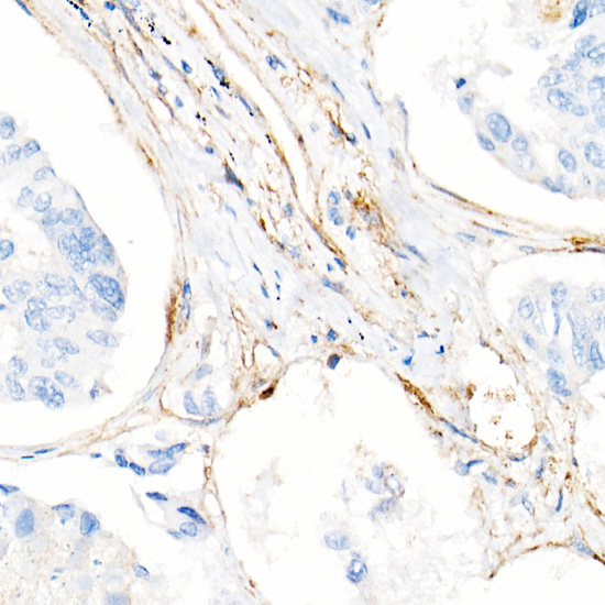
Immunohistochemistry analysis of paraffin-embedded Human esophageal cancer using Integrin-beta1/CD29 Rabbit mAb (A23497) at dilution of 1:5000 (40x lens). High pressure antigen retrieval performed with 0.01M Citrate buffer (pH 6.0) prior to IHC staining. |
|
|
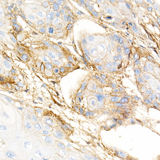
Immunohistochemistry analysis of paraffin-embedded Human lung cancer using Integrin-beta1/CD29 Rabbit mAb (A23497) at dilution of 1:5000 (40x lens). High pressure antigen retrieval performed with 0.01M Citrate buffer (pH 6.0) prior to IHC staining. |
|
|
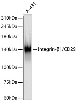
Western blot analysis of lysates from A-431 cells using Integrin-beta1/CD29 Rabbit mAb (A23497) at 1:22000 dilution incubated overnight at 4°C. Secondary antibody: HRP-conjugated Goat anti-Rabbit IgG (H+L) (AS014) at 1:10000 dilution. Lysates/proteins: 25 µg per lane. Blocking buffer: 3% nonfat dry milk in TBST. Detection: ECL Basic Kit (RM00020). Exposure time: 30s. |
|
|
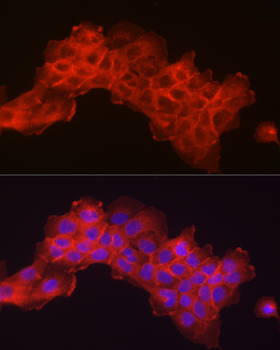
Immunofluorescence analysis of A-431 cells using Integrin-beta1/CD29 Rabbit mAb (A23497) at dilution of 1:100 (40x lens). Secondary antibody: Cy3-conjugated Goat anti-Rabbit IgG (H+L) (AS007) at 1:500 dilution. Blue: DAPI for nuclear staining. |
|
|
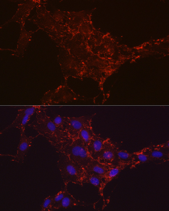
Immunofluorescence analysis of C6 cells using Integrin-beta1/CD29 Rabbit mAb (A23497) at dilution of 1:100 (40x lens). Secondary antibody: Cy3-conjugated Goat anti-Rabbit IgG (H+L) (AS007) at 1:500 dilution. Blue: DAPI for nuclear staining. |
|
|
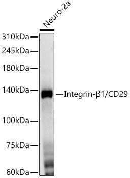
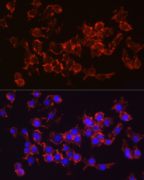
Immunofluorescence analysis of Neuro-2a cells using Integrin-beta1/CD29 Rabbit mAb (A23497) at dilution of 1:100 (40x lens). Secondary antibody: Cy3-conjugated Goat anti-Rabbit IgG (H+L) (AS007) at 1:500 dilution. Blue: DAPI for nuclear staining. |
|
|
Flow cytometry: 1X10 6 HL-60 cells (Low Expression,left)and A549 cells (right) were surface-stained with Rabbit anti-Human Integrin-beta1/CD29 mAb (A27614,2 µg/mL,orange line) or Rabbit IgG isotype control (AC042,2 µg/mL,blue line), followed by Alexa Fluor 647 conjugated goat anti-rabbit pAb staining. Non-fluorescently stained cells were used as blank control (red line). |
|
|
Flow cytometry: 1X10 6 A549 cells were surface-stained with Rabbit IgG isotype control (AC042,2 µg/mL,left) or Rabbit anti-Human Integrin-beta1/CD29 mAb (A27614,2 µg/mL,right), followed by Alexa Fluor 647 conjugated goat anti-rabbit pAb staining. |
|
|
Flow cytometry: 1X10 6 A549 cells were surface-stained with Rabbit IgG isotype control (AC042,2 µg/mL,left) or Rabbit anti-Human Integrin-beta1/CD29 mAb (A27614,2 µg/mL,right), followed by Alexa Fluor 647 conjugated goat anti-rabbit pAb staining. |
Produktgarantie und fachkundiger Support
