Enables several functions, including heparan sulfate proteoglycan binding activity, heparin binding activity, and protein tyrosine phosphatase activity. Involved in several processes, including lymphocyte differentiation, positive regulation of macromolecule metabolic process, and regulation of signal transduction. Acts upstream of or within several processes, including lymphocyte differentiation, positive regulation of lymphocyte activation, and regulation of protein phosphorylation. Located in external side of plasma membrane, focal adhesion, and membrane raft. Is expressed in several structures, including 3rd branchial arch, alimentary system, cardiovascular system, hemolymphoid system, and placenta. Used to study systemic lupus erythematosus. Human ortholog(s) of this gene implicated in hepatitis C, multiple sclerosis, severe combined immunodeficiency, and severe combined immunodeficiency, autosomal recessive, T cell-negative, B cell-positive, Nk cell-positive. Orthologous to human PTPRC (protein tyrosine phosphatase receptor type C).
IF/ICC,1:100 - 1:800|IF-F,1:100 - 1:800|IHC-P,1:500 - 1:2000|FC,1:100 - 1:500|ELISA,Recommended starting concentration is 1 µg/mL. Please optimize the concentration based on your specific assay requirements.
Anwendungsbeschreibung:
Cross-Reactivity: Mouse. ResearchArea: Immunology,Adaptive Immunity,B Cells,CD. Shipping: Ice Bag
Immunohistochemistry analysis of paraffin-embedded Mouse brain tissue using CD45 Rabbit mAb (A23549) at a dilution of 1:1000 (40x lens). High pressure antigen retrieval performed with 0.01M Tris-EDTA Buffer (pH 9.0) prior to IHC staining.
Immunohistochemistry analysis of paraffin-embedded Mouse intestine tissue using CD45 Rabbit mAb (A23549) at a dilution of 1:1000 (40x lens). High pressure antigen retrieval performed with 0.01M Tris-EDTA Buffer (pH 9.0) prior to IHC staining.
Immunohistochemistry analysis of paraffin-embedded Mouse liver tissue using CD45 Rabbit mAb (A23549) at a dilution of 1:1000 (40x lens). High pressure antigen retrieval performed with 0.01M Tris-EDTA Buffer (pH 9.0) prior to IHC staining.
Immunohistochemistry analysis of paraffin-embedded Mouse lung tissue using CD45 Rabbit mAb (A23549) at a dilution of 1:1000 (40x lens). High pressure antigen retrieval performed with 0.01M Tris-EDTA Buffer (pH 9.0) prior to IHC staining.
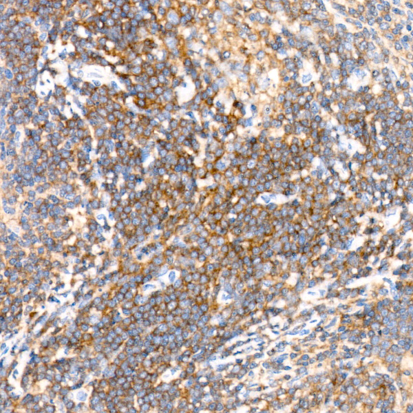
Immunohistochemistry analysis of paraffin-embedded Mouse spleen tissue using CD45 Rabbit mAb (A23549) at a dilution of 1:1000 (40x lens). High pressure antigen retrieval performed with 0.01M Tris-EDTA Buffer (pH 9.0) prior to IHC staining.
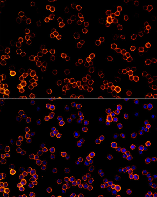
Immunofluorescence analysis of RAW264.7 cells using CD45 Rabbit mAb (A23549) at dilution of 1:100 (40x lens). Secondary antibody: Cy3-conjugated Goat anti-Rabbit IgG (H+L) (AS007) at 1:500 dilution. Blue: DAPI for nuclear staining.
Confocal imaging of frozen sections Mouse spleen tissue using CD45 Rabbit mAb (A23549, dilution 1:200) followed by a further incubation with Cy3 Goat Anti-Rabbit IgG (H+L) (AS007, dilution 1:500) (Red). DAPI was used for nuclear staining (Blue). Microwave antigen retrieval performed with 0.01M Citrate Buffer (pH 6.0) prior to IF staining. Objective: 40x.
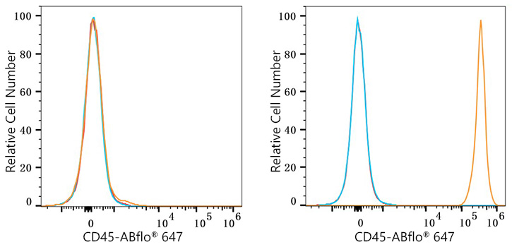
Flow cytometry: 1X10 6 C2C12 cells (negative control,left)and C57BL/6 mouse Splenocytes (right) were surface-stained with CD45 Rabbit mAb (A23549,2 µg/mL,orange line) or ABflo 647 Rabbit IgG isotype control (A22070,5 µl/Test,blue line), followed by Alexa Fluor 647 conjugated goat anti-rabbit pAb staining. Non-fluorescently stained cells were used as blank control (red line).
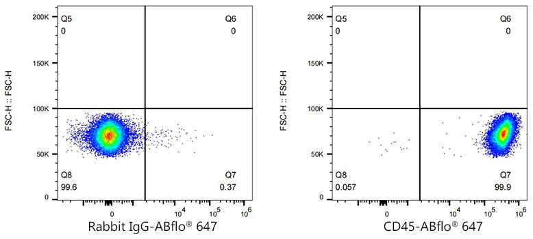
Flow cytometry: 1X10 6 C57BL/6 mouse Splenocytes were surface-stained with ABflo 647 Rabbit IgG isotype control (A22070,5 µl/Test,left) or CD45 Rabbit mAb (A23549,2 µg/mL,right).
* Mehrwertsteuer und Versandkosten nicht enthalten. Irrtümer und Preisänderungen vorbehalten