
[KD Validated] RAB27A Rabbit mAb, Unconjugated, Monoclonal
Artikelnummer:
ABB-A23993
- Bilder (9)


| Artikelname: | [KD Validated] RAB27A Rabbit mAb, Unconjugated, Monoclonal |
| Artikelnummer: | ABB-A23993 |
| Hersteller Artikelnummer: | A23993 |
| Alternativnummer: | ABB-A23993-1000UL,ABB-A23993-20UL,ABB-A23993-500UL,ABB-A23993-100UL |
| Hersteller: | ABclonal |
| Wirt: | Rabbit |
| Kategorie: | Antikörper |
| Applikation: | ELISA, IF, IHC-P, WB |
| Spezies Reaktivität: | Human |
| Immunogen: | Recombinant protein (or fragment).This information is considered to be commercially sensitive. |
| Konjugation: | Unconjugated |
| Alternative Synonym: | GS2, RAM, RAB27, HsT18676, RAB27A |
| The protein encoded by this gene belongs to the small GTPase superfamily, Rab family. The protein is membrane-bound and may be involved in protein transport and small GTPase mediated signal transduction. Mutations in this gene are associated with Griscelli syndrome type 2. Alternative splicing occurs at this locus and four transcript variants encoding the same protein have been identified. |
| Klonalität: | Monoclonal |
| Klon-Bezeichnung: | [ARC61834] |
| Molekulargewicht: | 25kDa |
| NCBI: | 5873 |
| UniProt: | P51159 |
| Reinheit: | Affinity purification |
| Sequenz: | MSDGDYDYLIKFLALGDSGVGKTSVLYQYTDGKFNSKFITTVGIDFREKRVVYRASGPDGATGRGQRIHLQLWDTAGQERFRSLTTAFFRDAMGFLLLFDLTNEQSFLNVRNWISQLQMHAYCENPDIVLCGNKSDLEDQRVVKEEEAIALAEKYGIPYFETSAANGTNISQAIEMLLDLIMKRMERCVDKSWIPEGVVRSNGHASTDQLSEEKEKGACGC |
| Target-Kategorie: | RAB27A |
| Antibody Type: | Primary Antibody |
| Application Verdünnung: | WB,1:1500 - 1:6000|IF/ICC,1:200 - 1:800|IF-P,1:200 - 1:800|IHC-P,1:200 - 1:2000|ELISA,Recommended starting concentration is 1 µg/mL. Please optimize the concentration based on your specific assay requirements. |
| Anwendungsbeschreibung: | Cross-Reactivity: Human,Mouse,Rat. ResearchArea: Signal Transduction,G protein signaling,Small G proteins,Cell Biology Developmental Biology,Apoptosis. Shipping: Ice Bag |
|
|
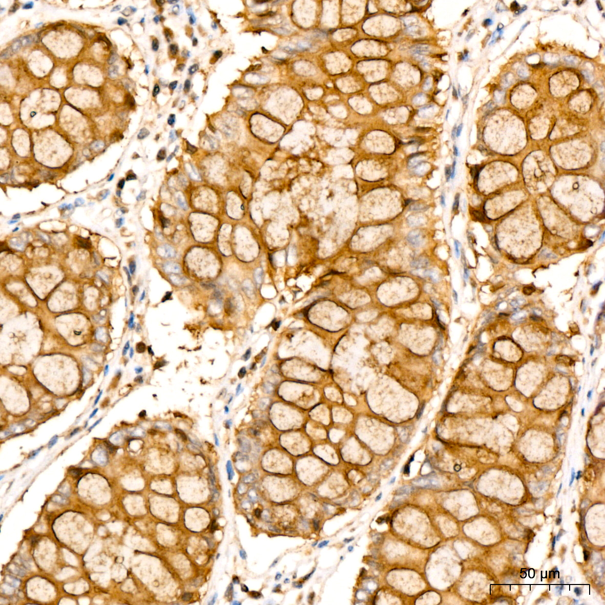
Immunohistochemistry analysis of paraffin-embedded Human colon carcinoma tissue using [KD Validated] RAB27A Rabbit mAb (A23993) at dilution of 1:300 (40x lens). High pressure antigen retrieval performed with 0.01M Citrate Buffer (pH 6.0) prior to IHC staining. |
|
|
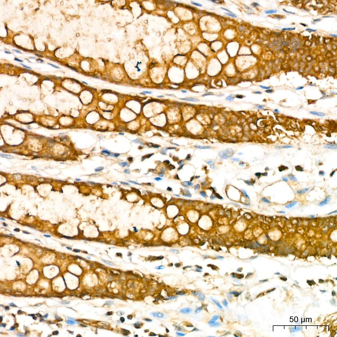
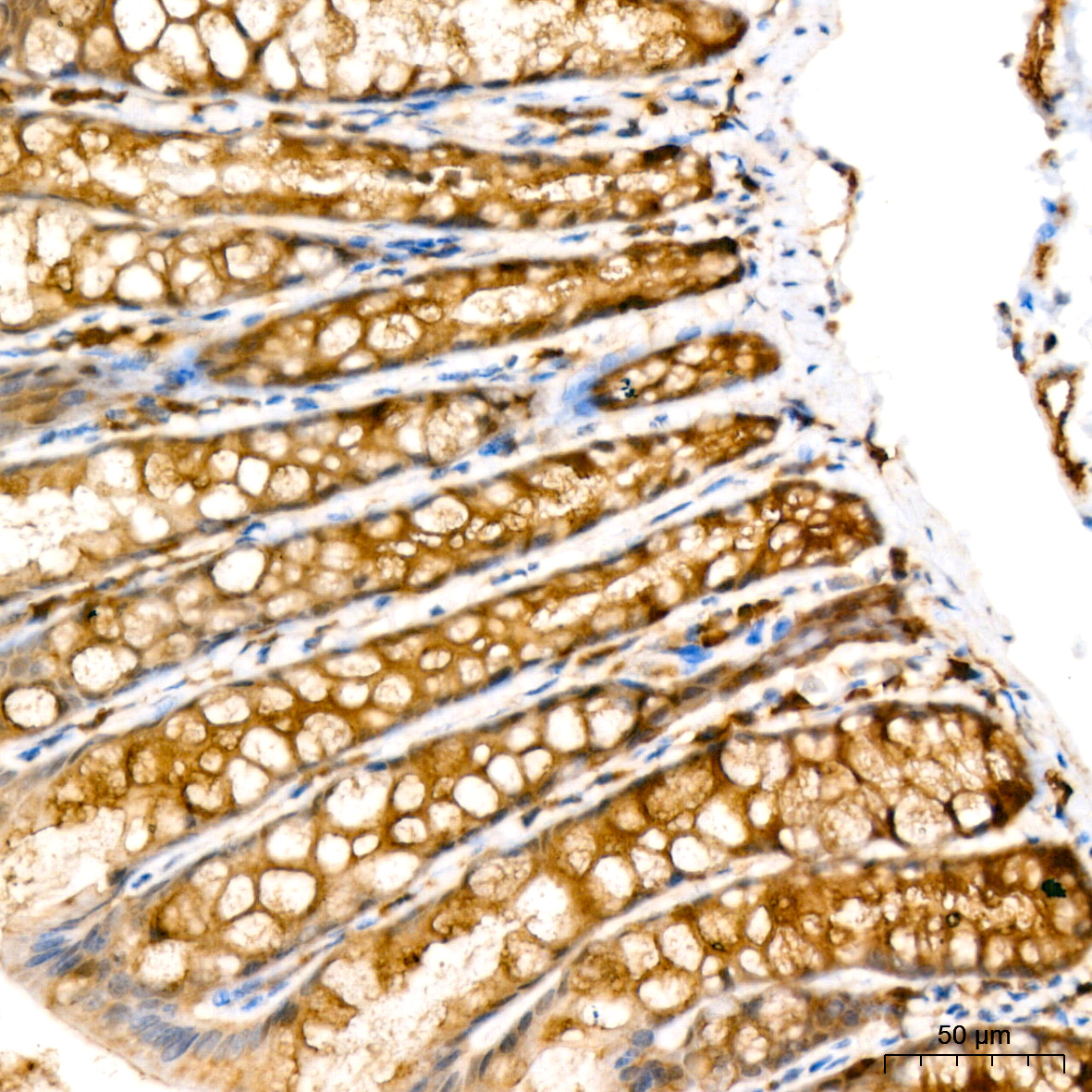
Immunohistochemistry analysis of paraffin-embedded Human colon tissue using [KD Validated] RAB27A Rabbit mAb (A23993) at dilution of 1:300 (40x lens). High pressure antigen retrieval performed with 0.01M Citrate Buffer (pH 6.0) prior to IHC staining. |
|
|
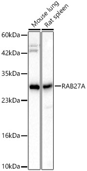
Western blot analysis of various lysates, using [KD Validated] RAB27A Rabbit mAb (A23993) at 1:3000 dilution. Secondary antibody: HRP-conjugated Goat anti-Rabbit IgG (H+L) (AS014) at 1:10000 dilution. Lysates/proteins: 25µg per lane. Blocking buffer: 3% nonfat dry milk in TBST. Detection: ECL Basic Kit (RM00020). Exposure time: 45s. |
|
|
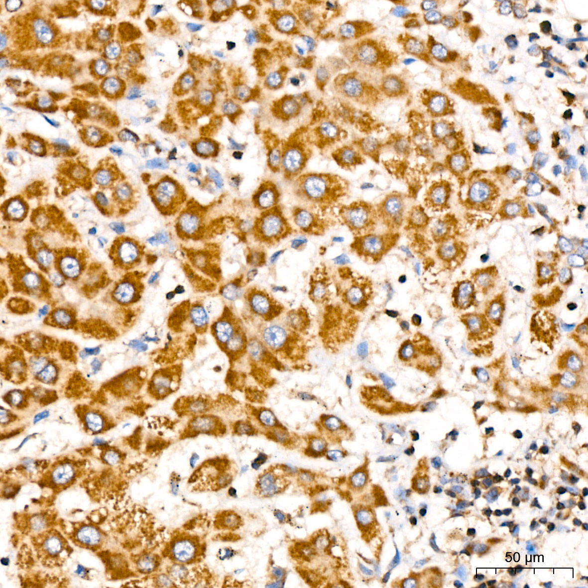
Immunohistochemistry analysis of paraffin-embedded Human liver tissue using [KD Validated] RAB27A Rabbit mAb (A23993) at dilution of 1:300 (40x lens). High pressure antigen retrieval performed with 0.01M Citrate Buffer (pH 6.0) prior to IHC staining. |
|
|
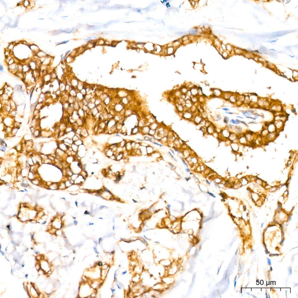
Immunohistochemistry analysis of paraffin-embedded Human thyroid cancer tissue using [KD Validated] RAB27A Rabbit mAb (A23993) at dilution of 1:300 (40x lens). High pressure antigen retrieval performed with 0.01M Citrate Buffer (pH 6.0) prior to IHC staining. |
|
|
Immunohistochemistry analysis of paraffin-embedded Mouse colon tissue using [KD Validated] RAB27A Rabbit mAb (A23993) at dilution of 1:300 (40x lens). High pressure antigen retrieval performed with 0.01M Citrate Buffer (pH 6.0) prior to IHC staining. |
|
|
Confocal imaging of Hep G2 cells using [KD Validated] RAB27A Rabbit mAb (A23993, dilution 1:200) followed by a further incubation with Cy3 Goat Anti-Rabbit IgG (H+L) (AS007, dilution 1:500) (Red). The cells were counterstained with alpha-tubulin Mouse mAb (AC012, dilution 1:400) followed by incubation with ABflo 488-conjugated Goat Anti-Mouse IgG (H+L) Ab (AS076, dilution 1:500) (Green). DAPI was used for nuclear staining (Blue). Objective: 100x. |
|
|
Confocal imaging of paraffin-embedded Mouse colon tissue using [KD Validated] RAB27A Rabbit mAb (A23993, dilution 1:200) followed by a further incubation with Cy3 Goat Anti-Rabbit IgG (H+L) (AS007, dilution 1:500) (Red). DAPI was used for nuclear staining (Blue). High pressure antigen retrieval performed with 0.01M Citrate Buffer(pH 6.0) prior to IF staining. Objective: 40x. |
|
|
Confocal imaging of PC-12 cells using [KD Validated] RAB27A Rabbit mAb (A23993, dilution 1:200) followed by a further incubation with Cy3 Goat Anti-Rabbit IgG (H+L) (AS007, dilution 1:500) (Red). The cells were counterstained with alpha-Tubulin Mouse mAb (AC012, dilution 1:400) followed by incubation with ABflo 488-conjugated Goat Anti-Mouse IgG (H+L) Ab (AS076, dilution 1:500) (Green). DAPI was used for nuclear staining (Blue). Objective: 100x. |
Produktgarantie und fachkundiger Support
