
ACE2 Rabbit mAb, Unconjugated, Monoclonal
Artikelnummer:
ABB-A4612
- Bilder (6)
| Artikelname: | ACE2 Rabbit mAb, Unconjugated, Monoclonal |
| Artikelnummer: | ABB-A4612 |
| Hersteller Artikelnummer: | A4612 |
| Alternativnummer: | ABB-A4612-20UL,ABB-A4612-100UL,ABB-A4612-1000UL,ABB-A4612-500UL |
| Hersteller: | ABclonal |
| Wirt: | Rabbit |
| Kategorie: | Antikörper |
| Applikation: | ELISA, IF, IHC-P, WB |
| Spezies Reaktivität: | Human |
| Immunogen: | Recombinant protein (or fragment).This information is considered to be commercially sensitive. |
| Konjugation: | Unconjugated |
| Alternative Synonym: | ACEH, ACE2 |
| The protein encoded by this gene belongs to the angiotensin-converting enzyme family of dipeptidyl carboxydipeptidases and has considerable homology to human angiotensin 1 converting enzyme. This secreted protein catalyzes the cleavage of angiotensin I into angiotensin 1-9, and angiotensin II into the vasodilator angiotensin 1-7. ACE2 is known to be expressed in various human organs, and its organ- and cell-specific expression suggests that it may play a role in the regulation of cardiovascular and renal function, as well as fertility. In addition, the encoded protein is a functional receptor for the spike glycoprotein of the human coronavirus HCoV-NL63 and the human severe acute respiratory syndrome coronaviruses, SARS-CoV and SARS-CoV-2, the latter is the causative agent of coronavirus disease-2019 (COVID-19). Multiple splice variants have been found for this gene and the dACE2 (or MIRb-ACE2) splice variant has been found to be interferon inducible. |
| Klonalität: | Monoclonal |
| Klon-Bezeichnung: | [ARC50948] |
| Molekulargewicht: | 92 kDa |
| NCBI: | 59272 |
| UniProt: | Q9BYF1 |
| Reinheit: | Affinity purification |
| Sequenz: | QSTIEEQAKTFLDKFNHEAEDLFYQSSLASWNYNTNITEENVQNMNNAGDKWSAFLKEQSTLAQMYPLQEIQNLTVKLQLQALQQNGSSVLSEDKSKRLNTILNTMSTIYSTGKVCNPDNPQECLLLEPGLNEIMANSLDYNERLWAWESWRSEVGKQLRPLYEEYVVLKNEMARANHYEDYGDYWRGDYEVNGVDGYDYSRGQLIEDVEHTFEEIKPLYEHLHAYVRAKLMNAYPSYISPIGCLPAHLLGDMWG |
| Target-Kategorie: | ACE2 |
| Antibody Type: | Primary Antibody |
| Application Verdünnung: | WB,1:1000 - 1:5000|IF-P,1:100 - 1:1000|IHC-P,1:200 - 1:2000|ELISA,Recommended starting concentration is 1 µg/mL. Please optimize the concentration based on your specific assay requirements. |
| Anwendungsbeschreibung: | Cross-Reactivity: Human,Mouse,Rat. ResearchArea: Cell Biology Developmental Biology,Ubiquitin,Cardiovascular,Blood,Heart,Contractility. Shipping: Ice Bag |
|
|
Western blot analysis of lysates from Rat kidney using ACE2 Rabbit mAb (A4612) at 1:4000 dilution incubated at room temperature for 1.5 hours. Secondary antibody: HRP-conjugated Goat anti-Rabbit IgG (H+L) (AS014) at 1:10000 dilution. Lysates/proteins: 25 µg per lane. Blocking buffer: 3% nonfat dry milk in TBST. Detection: ECL Basic Kit (RM00020). Exposure time: 90 s. |
|
|
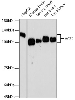
Western blot analysis of various lysates using ACE2 Rabbit mAb (A4612) at 1:5000 dilutionincubated overnight at 4°C. Secondary antibody: HRP-conjugated Goat anti-Rabbit IgG (H+L) (AS014) at 1:10000 dilution. Lysates/proteins: 25 µg per lane. Blocking buffer: 3% nonfat dry milk in TBST. Detection: ECL Basic Kit (RM00020). Negative control (NC): Hep G2, HeLa. Exposure time: 90 s. |
|
|
Western blot analysis of lysates from Mouse kidney using ACE2 Rabbit mAb (A4612) at 1:4000 dilution incubated at room temperature for 1.5 hours. Secondary antibody: HRP-conjugated Goat anti-Rabbit IgG (H+L) (AS014) at 1:10000 dilution. Lysates/proteins: 25 µg per lane. Blocking buffer: 3% nonfat dry milk in TBST. Detection: ECL Basic Kit (RM00020). Exposure time: 60 s. |
|
|
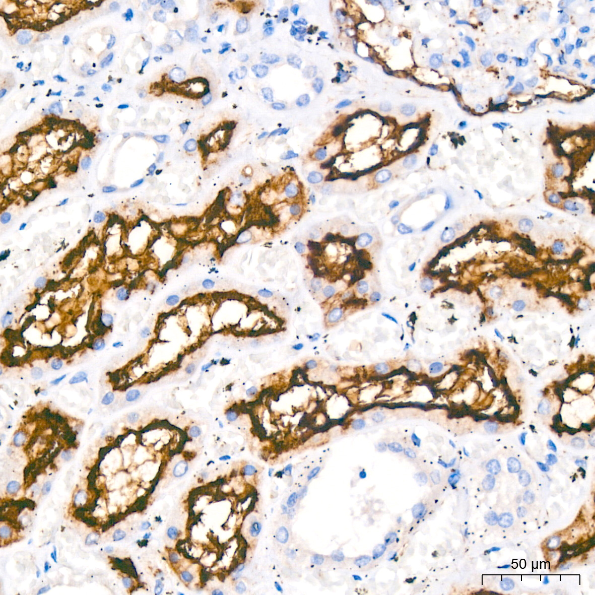
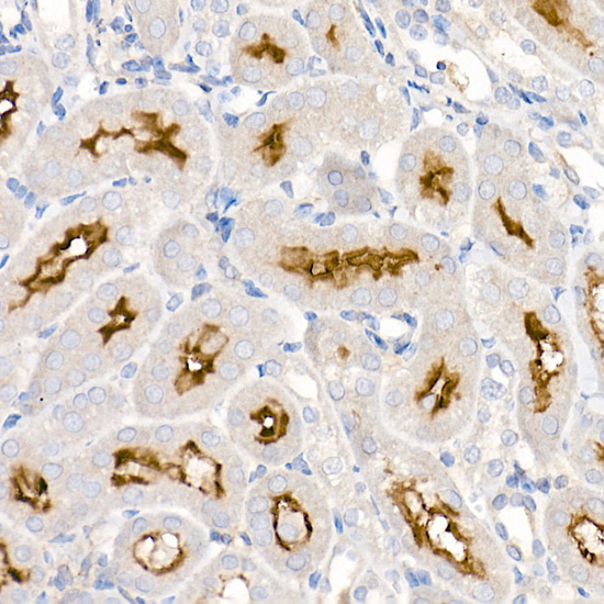
Immunohistochemistry analysis of paraffin-embedded Human kidney using ACE2 Rabbit mAb (A4612) at dilution of 1:200 (40x lens). High pressure antigen retrieval performed with 0.01M Citrate buffer (pH 6.0) prior to IHC staining. |
|
|
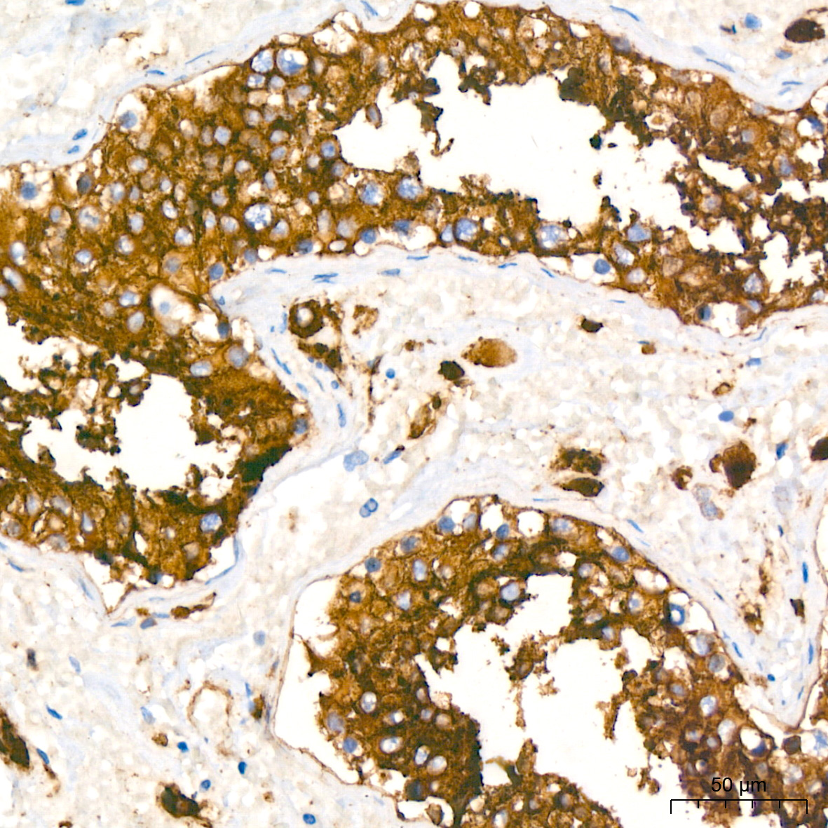
Immunohistochemistry analysis of paraffin-embedded Human testis using ACE2 Rabbit mAb (A4612) at dilution of 1:200 (40x lens). High pressure antigen retrieval performed with 0.01M Citrate buffer (pH 6.0) prior to IHC staining. |
|
|
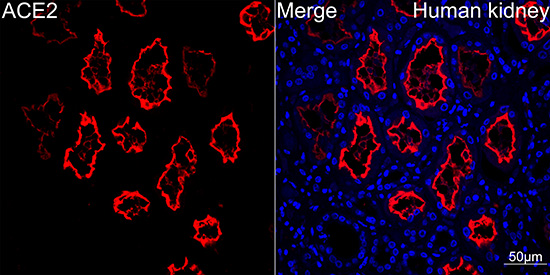
Confocal imaging of Human kidney using ACE2 Rabbit mAb (A4612,dilution 1:100)(Red). DAPI was used for nuclear staining (blue). Objective: 40x. |
Produktgarantie und fachkundiger Support
