
Phospho-JNK1-T183/Y185 + JNK2-T183/Y185 + JNK3-T221/Y223 Rabbit pAb
Artikelnummer:
ABB-AP1163
- Bilder (1)
| Artikelname: | Phospho-JNK1-T183/Y185 + JNK2-T183/Y185 + JNK3-T221/Y223 Rabbit pAb |
| Artikelnummer: | ABB-AP1163 |
| Hersteller Artikelnummer: | AP1163 |
| Alternativnummer: | ABB-AP1163-100UL, ABB-AP1163-20UL |
| Hersteller: | ABclonal |
| Wirt: | Rabbit |
| Kategorie: | Antikörper |
| Applikation: | ELISA, WB |
| Spezies Reaktivität: | Human |
| Immunogen: | Synthetic peptide. This information is considered to be commercially sensitive. |
| The protein encoded by this gene is a member of the MAP kinase family. MAP kinases act as an integration point for multiple biochemical signals, and are involved in a wide variety of cellular processes such as proliferation, differentiation, transcription regulation and development. This kinase is activated by various cell stimuli, and targets specific transcription factors, and thus mediates immediate-early gene expression in response to cell stimuli. The activation of this kinase by tumor-necrosis factor alpha (TNF-alpha) is found to be required for TNF-alpha induced apoptosis. This kinase is also involved in UV radiation induced apoptosis, which is thought to be related to cytochrom c-mediated cell death pathway. Studies of the mouse counterpart of this gene suggested that this kinase play a key role in T cell proliferation, apoptosis and differentiation. Several alternatively spliced transcript variants encoding distinct isoforms have been reported. [provided by RefSeq, Apr 2016] |
| Application Verdünnung: | WB,1:500 - 1:1000|ELISA,Recommended starting concentration is 1 µg/mL. Please optimize the concentration based on your specific assay requirements. |
| Anwendungsbeschreibung: | Cross-Reactivity: Human,Mouse |
|
|
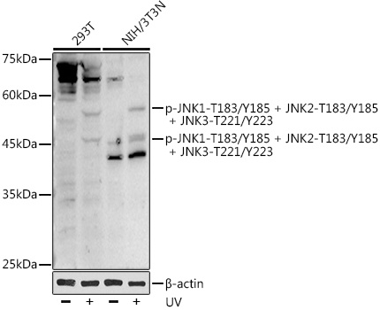
Western blot analysis of various lysates using Phospho-JNK1-T183/Y185 + JNK2-T183/Y185 + JNK3-T221/Y223 Rabbit pAb (AP1163) at 1:1000 dilution. 293T cells were treated with UV at room temperature for 15-30 minutes. NIH/3T3 cells were treated with UV at room temperature for 15-30 minutes. Secondary antibody: HRP-conjugated Goat anti-Rabbit IgG (H+L) (AS014) at 1:10000 dilution. Lysates/proteins: 25µg per lane. Blocking buffer: 3% nonfat dry milk in TBST. Detection: ECL Basic Kit (RM00020). Exposure time: 180s. |
Produktgarantie und fachkundiger Support
