
Phospho-FAK-Y397 Rabbit mAb, Unconjugated
Artikelnummer:
ABB-AP1447
- Bilder (1)
| Artikelname: | Phospho-FAK-Y397 Rabbit mAb, Unconjugated |
| Artikelnummer: | ABB-AP1447 |
| Hersteller Artikelnummer: | AP1447 |
| Alternativnummer: | ABB-AP1447-20UL,ABB-AP1447-100UL,ABB-AP1447-1000UL,ABB-AP1447-500UL |
| Hersteller: | ABclonal |
| Wirt: | Rabbit |
| Kategorie: | Antikörper |
| Applikation: | ELISA, WB |
| Spezies Reaktivität: | Human |
| Immunogen: | Synthetic peptide. This information is considered to be commercially sensitive. |
| Konjugation: | Unconjugated |
| Alternative Synonym: | FAK, FADK, FAK1, FRNK, FADK 1, PPP1R71, p125FAK, pp125FAK, Phospho-FAK-Y397 |
| This gene encodes a cytoplasmic protein tyrosine kinase which is found concentrated in the focal adhesions that form between cells growing in the presence of extracellular matrix constituents. The encoded protein is a member of the FAK subfamily of protein tyrosine kinases but lacks significant sequence similarity to kinases from other subfamilies. Activation of this gene may be an important early step in cell growth and intracellular signal transduction pathways triggered in response to certain neural peptides or to cell interactions with the extracellular matrix. Several transcript variants encoding different isoforms have been found for this gene. |
| Application Verdünnung: | WB,1:2000 - 1:4000|ELISA,Recommended starting concentration is 1 µg/mL. Please optimize the concentration based on your specific assay requirements. |
| Anwendungsbeschreibung: | Cross-Reactivity: Human, ResearchArea: Protein phosphorylation,Cancer,Signal Transduction,G protein signaling,G-Protein-Coupled Receptors Signaling to MAPK Erk,Kinase,Tyrosine kinases,PI3K-Akt Signaling Pathway,ErbB-HER Signaling Pathway,MAPK-Erk Signaling Pathway,Cell Biology Developmental Biology,Apoptosis,Cytoskeleton,Actins,Extracellular Matrix,Immunology Inflammation,Cardiovascular,Angiogenesis. |
|
|
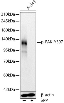
Western blot analysis of lysates from A-549 cells, using Phospho-FAK-Y397 Rabbit mAb (AP1447) at 1:4000 dilution. A-549 cells were treated by lambda-PP mixed solution (1ul) at 30°C for 30 minutes. Secondary antibody: HRP-conjugated Goat anti-Rabbit IgG (H+L) (AS014) at 1:10000 dilution. Lysates/proteins: 25µg per lane. Blocking buffer: 3% nonfat dry milk in TBST. Detection: ECL Basic Kit (RM00020). Exposure time: 30s. |
Produktgarantie und fachkundiger Support
