Specificity : Recognizes human ACE2. Angiotensin-converting enzyme2 (ACE2) is an ectoenzyme (carboxypeptidase) with an extracellular catalytic domain that predominantly localizes at the plasma membrane and is thereby able to hydrolyze circulating peptides. ACE2 has approximately 42% sequence identity with ACE, and its cytoplasmic and transmembrane domains show 48% homology to the protein collectrin that plays a critical role in the amino acid absorption of the kidney. ACE2 converts angiotensin I to angiotensin 1-9, a peptide of unknown function, and angiotensin II to angiotensin 1-7, a vasodilator. ACE2 is involved in the regulation of systemic blood pressure and has direct effects on cardiac functions. It is expressed predominantly in endothelial cells of the lung, gut, heart and kidney. ACE2 together with the protease TMPRSS2 acts as a functional receptor for SARS coronavirus as well as for the new highly pathogenic coronavirus, 2019-nCoV/SARS-CoV-2, which is cause for pneumonia COVID-19.
Flow Cytometry: (1:1000) Optimal conditions should be determined individually for each application.
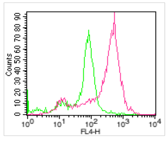
Fig. 2: Cell Surface staining of ACE2 on PBMC (lymphocytes). Red: Atto 647 conjugated human anti-ACE2 antibody (clone AC18F) (1ug/10 6 cells) were used. Green: Isotype control mouse IgG1 conjugated with Atto 647 (1ug/ 10 6 cells) was used as control.
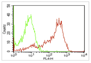
Fig.1: Cell Surface staining of ACE2 on HepG2 cell line. Red: Atto 647 conjugated human anti-ACE2 antibody (clone AC18F) (1ug/10 6 cells) were used. Green: Atto 647 conjugated Isotype control, mouse IgG1 A (1ug/ 10 6 cells) was used as control.
* Mehrwertsteuer und Versandkosten nicht enthalten. Irrtümer und Preisänderungen vorbehalten